2. Course _Protozoa_Engl (1).pdf - english section
1.
U.A.S.V.M. of Bucharest
Facultyof Veterinary Medicine
Department of Preclinical Sciences
Discipline: Animal Biology
Course
Assoc. Prof. PhD Mariana IONITA
Habilitated Doctor, Dipl. EVPC
2.
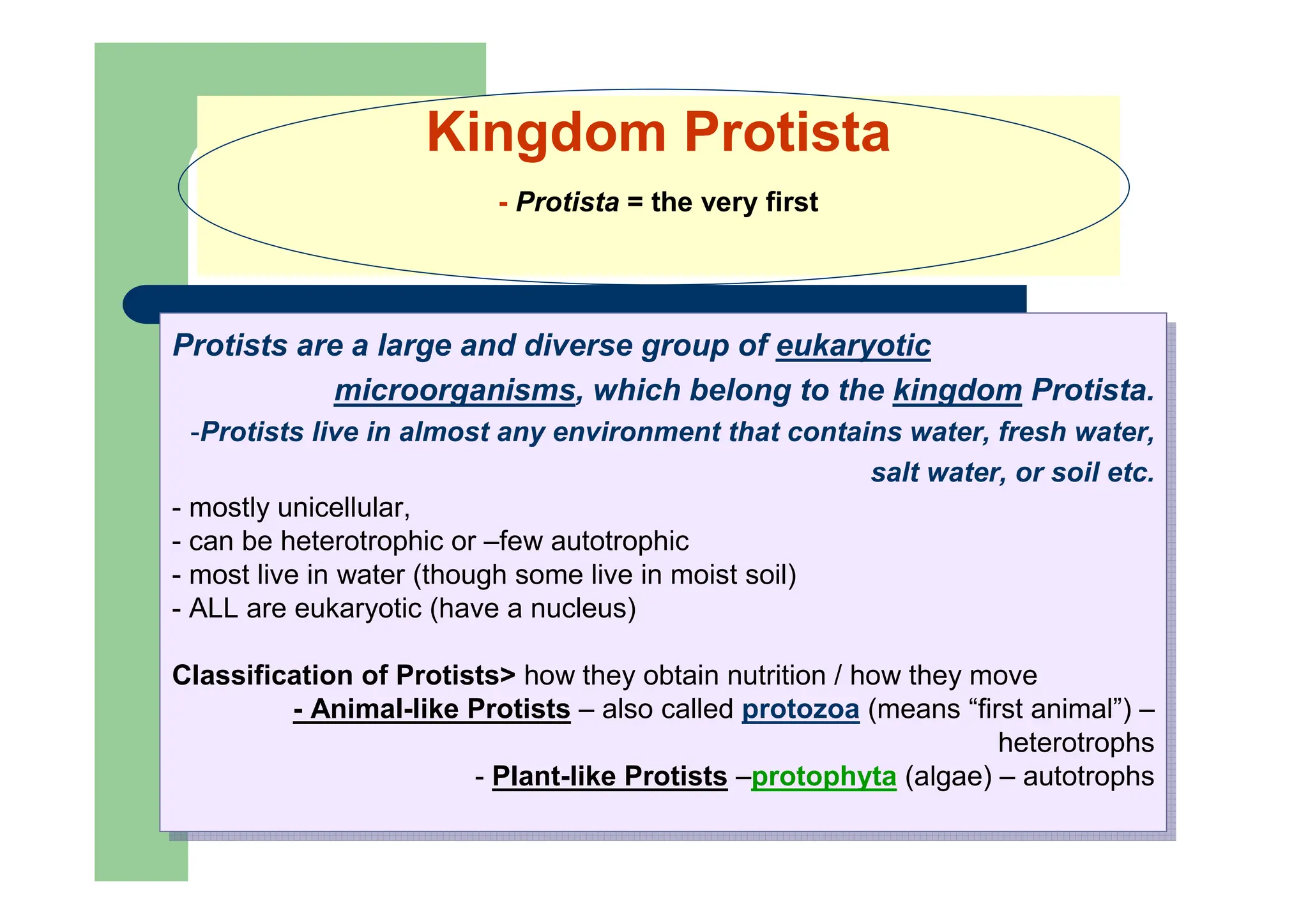
Kingdom Protista
- Protista= the very first
Protists are a large and diverse group of eukaryotic
microorganisms, which belong to the kingdom Protista.
-Protists live in almost any environment that contains water, fresh water,
salt water, or soil etc.
- mostly unicellular,
- can be heterotrophic or –few autotrophic
- most live in water (though some live in moist soil)
- ALL are eukaryotic (have a nucleus)
Classification of Protists> how they obtain nutrition / how they move
- Animal-like Protists – also called protozoa (means “first animal”) –
heterotrophs
- Plant-like Protists –protophyta (algae) – autotrophs
3.
Protozoa are adiverse group of unicellular eukaryotic organisms,
- usually single-celled and heterotrophic eukaryotes containing non-
filamentous structures that belong to any of the major lineages of protists.
- They are restricted to moist or aquatic habitats:
-i.e., they are obligate aquatic organisms;
- many protozoan species are symbionts,
- some are parasites,
- and some are predators of bacteria and algae.
There are an estimated 30,000 protozoan species]
Protozoa are a diverse group of unicellular eukaryotic organisms,
- usually single-celled and heterotrophic eukaryotes containing non-
filamentous structures that belong to any of the major lineages of protists.
- They are restricted to moist or aquatic habitats:
-i.e., they are obligate aquatic organisms;
- many protozoan species are symbionts,
- some are parasites,
- and some are predators of bacteria and algae.
There are an estimated 30,000 protozoan species]
Phylum Protozoa Greek protos = first, zoon = animal)
I. Characteristics of protozoa
5.
• Protozoa commonlyrange in length between 10 to 52 micrometers,
but can grow as large as 1 mm.
• They are easily seen with a microscope.
• Protozoa exist throughout aqueous environments and soil, occupying
a range of trophic levels.
• protozoans: can be flagellates (motile with flagella), ciliates (motile
with cilia), and amoebas (motile by means of pseudopodia).
• Flagellates are the most numerous soil protozoa
Characteristics of protozoa
6.
Motility:
Some protoozoa canmove with:
– whip-like tails called flagella,
– hair-like structures called cilia, or
– foot-like structures called pseudopodia.
Others do not move at all.
Characteristics of protozoa
7.
Digestion
Protozoa may absorbfood via their cell
membranes,
some, e.g., amoebas, surround food and engulf it,
and
ingestion: others have openings or "mouth
pores" into which they sweep food,and that
engulfing of food is said to be phagocytosis.
All protozoa digest their food in compartments
called vacuoles (digestive vacuoles).
Characteristics of protozoa
8.
Protozoa: biology
Nutrition:
Parasitic protozoaby::
Osmosis
Phagocytosis: is the process by which a cell -often a
phagocyte or a protist—engulfs a solid particle to form an internal
compartment known as a phagosome.
Pinocitosis: involves the internalization of extracellular liquids.
some ciliated protozoa and also some stages of the organisms
causing different diseases (malaria) obtain food through a
cytostome:
At the base of the cytostome the food enters a vacuole for
digestion within the cell.
Metabolic products are excreted by diffusion through the cell
membrane.
9.
Life cycle ofprotozoa
Some protozoa have life stages alternating between proliferative
stages (e.g., trophozoites) and dormant cysts.
When protozoa are in the form of trophozoites (Greek, tropho = to
nourish), they actively feed.
As cysts, protozoa can survive harsh conditions, such as exposure to
extreme temperatures or harmful chemicals, or long periods without access to
nutrients, water, or oxygen for a period of time.
Being a cyst enables parasitic species to survive outside of a host,
and allows their transmission from one host to another.
The conversion of a trophozoite to cyst form is known as
encystation, while the process of transforming back into a trophozoite is
known as excystation.
Protozoa can Reproduce by binary fission or multiple fission.
Some protozoa reproduce sexually, some asexually,
while some use a combination, (e.g., Coccidia).
10.
Reproduction
Asexual reproduction:
Binary fission(equal binary fission): in which a cell divides into two
daughter cells (after the chromosomes have been duplicated and distributed
between them). This asexual mode of reproduction leads to rapid population
growth; It may be:
longitudinal
transverse
Internal budding:
Endodiogeny: a new organism develops from an outgrowth or bud due
to cell division at one particular site.
Endopoliogeny: many organisms..
Schizogony (merogony): multiple fission manifested either as:
merogony - results in merozoites: which are multiple daughter cells, that
originate within the same cell membrane:
trophozoite grows to a large size while the nucleus divides repeatedly.
This structure is called a meront (schizont) and, when mature, each
nucleus has acquired a portion of the cytoplasm so that the schizont
is filled with a large number of elongated separate organisms called
merozoites. The meront eventually ruptures, liberating the individual
merozoites.
sporogony results in sporozoites
[Sporogony follows sexual reproduction].
Protozoa: biology
11.
Reproduction
Sexual reproduction:
Gametogony:
Gamonts:
Microgamonts –producing microgametes
Macrogamonts – producing macrogamete.
fecundation - syngamy – results in zygote – egg-cell.
conjugation:
in some ciliates);
two cells form a bridge between their cytoplasm, the micronuclei
undergo meiosis, the macronuclei disappear, and the
haploid micronuclei are exchanged over the bridge
In most ciliate groups, however, the cells separate after conjugation,
and both form new macronuclei from their micronuclei.
Conjugation is followed by fission:
Protozoa: biology
12.
Reproduction
Both, asexual andsexual reproduction, by alternative
phases [in Apicomplexa protozoa]: the life cycle consists of:
Schizogony (asexual phase):
Gametogony (sexual phase)
Microgamonts – producing microgametes
Macrogamonts – producing macrogamete.
fecundation - syngamy – results in zygote – egg-cell.
Sporogony – results in oocysts (with sporozoites)
Example: Eimeria (Coccidia) sporulated oocysts have 4
sporoblasts, each with two sporozoites.
Example>
in Eimeria, both asexual and sexual phases occur in the same host
followed by a free-living phase – sporogony (in the environment,
outside of the host).
in others, such as Plasmodium / Babesia, the asexual phase occurs
in the vertebrate host and the sexual phase in the arthropod vector.
Protozoa: biology
13.
As components ofthe micro- and meiofauna, protozoa are an
important food source for microinvertebrates.
Thus, the ecological role of protozoa in the transfer of bacterial
and algal production to successive trophic levels is important.
As predators, they prey upon unicellular or filamentous algae,
bacteria, and microfungi.
Protozoa are consumers in the decomposer link of the food chain.
They also control bacteria populations and biomass to some extent.
On average, Protozoa eat ~ 100 to 1,000 bacteria per hour.
Protozoa can stimulate organic matter decomposition,
digest cellulose in rumen of cows and termite guts, and play a role in
nutrient mobilization.
Protozoa such as the malaria parasites (Plasmodium spp.),
trypanosomes and leishmania, coccidia, piroplasms, are also
important disease causing agents in humans and many other in
animals.
Ecological role of protozoa
14.
CLASSIFICATION /
taxonomy
The classificationof protozoa has been and remains a problematic
area of taxonomy.
Where they are available, DNA sequences are used as the basis
for classification but for the majority of described protozoa such
material is not available.
They have been and still are mostly on the basis of their
morphology and for the parasitic species their hosts.
Protozoa have been divided traditionally on the
basis of their means of locomotion.
15.
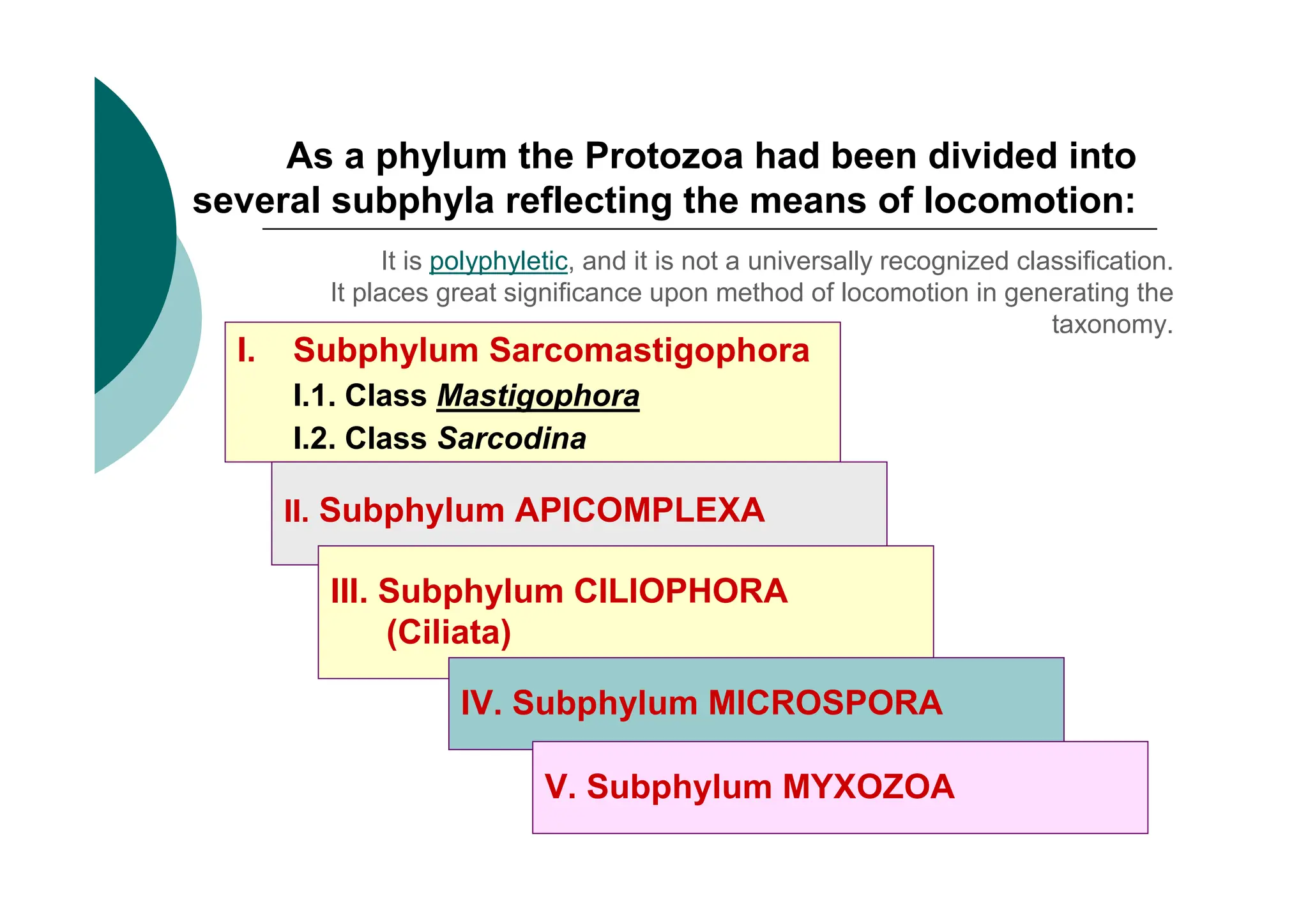
As a phylumthe Protozoa had been divided into
several subphyla reflecting the means of locomotion:
I. Subphylum Sarcomastigophora
I.1. Class Mastigophora
I.2. Class Sarcodina
It is polyphyletic, and it is not a universally recognized classification.
It places great significance upon method of locomotion in generating the
taxonomy.
II. Subphylum APICOMPLEXA
III. Subphylum CILIOPHORA
(Ciliata)
IV. Subphylum MICROSPORA
V. Subphylum MYXOZOA
16.
Phylum Protozoa –taxonomy ..ever changin
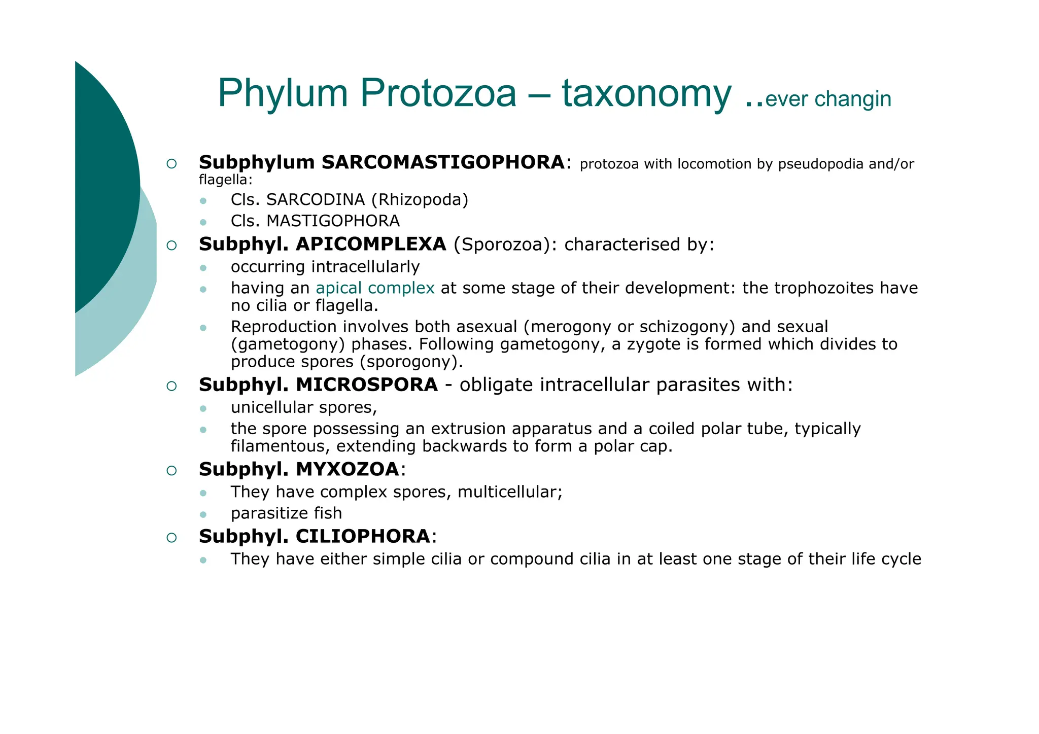
Subphylum SARCOMASTIGOPHORA: protozoa with locomotion by pseudopodia and/or
flagella:
Cls. SARCODINA (Rhizopoda)
Cls. MASTIGOPHORA
Subphyl. APICOMPLEXA (Sporozoa): characterised by:
occurring intracellularly
having an apical complex at some stage of their development: the trophozoites have
no cilia or flagella.
Reproduction involves both asexual (merogony or schizogony) and sexual
(gametogony) phases. Following gametogony, a zygote is formed which divides to
produce spores (sporogony).
Subphyl. MICROSPORA - obligate intracellular parasites with:
unicellular spores,
the spore possessing an extrusion apparatus and a coiled polar tube, typically
filamentous, extending backwards to form a polar cap.
Subphyl. MYXOZOA:
They have complex spores, multicellular;
parasitize fish
Subphyl. CILIOPHORA:
They have either simple cilia or compound cilia in at least one stage of their life cycle
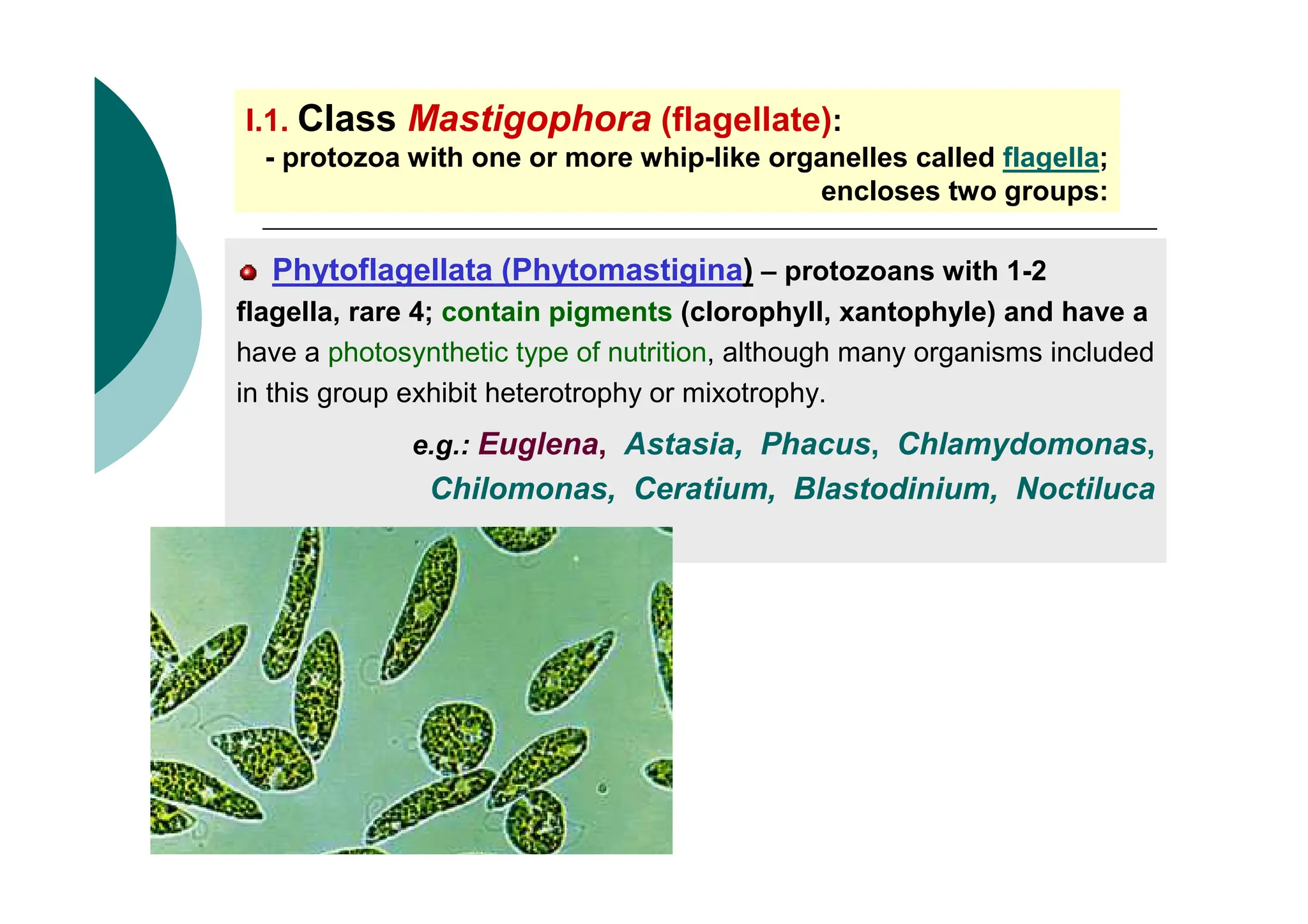
I.1. Class Mastigophora(flagellate):
- protozoa with one or more whip-like organelles called flagella;
encloses two groups:
Phytoflagellata (Phytomastigina) – protozoans with 1-2
flagella, rare 4; contain pigments (clorophyll, xantophyle) and have a
have a photosynthetic type of nutrition, although many organisms included
in this group exhibit heterotrophy or mixotrophy.
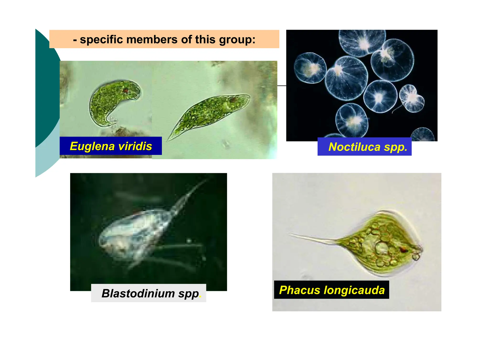
e.g.: Euglena, Astasia, Phacus, Chlamydomonas,
Chilomonas, Ceratium, Blastodinium, Noctiluca
I.1. Class Mastigophora
•group of Zooflagellata (Zoomastigina):
- protozoa with two or more flagella; some have pseudopodia;
- no color, heterotrophic (holozoice or saprozoice);
- some are free (eg coanoflagelatele); however, the majority are parasitic.
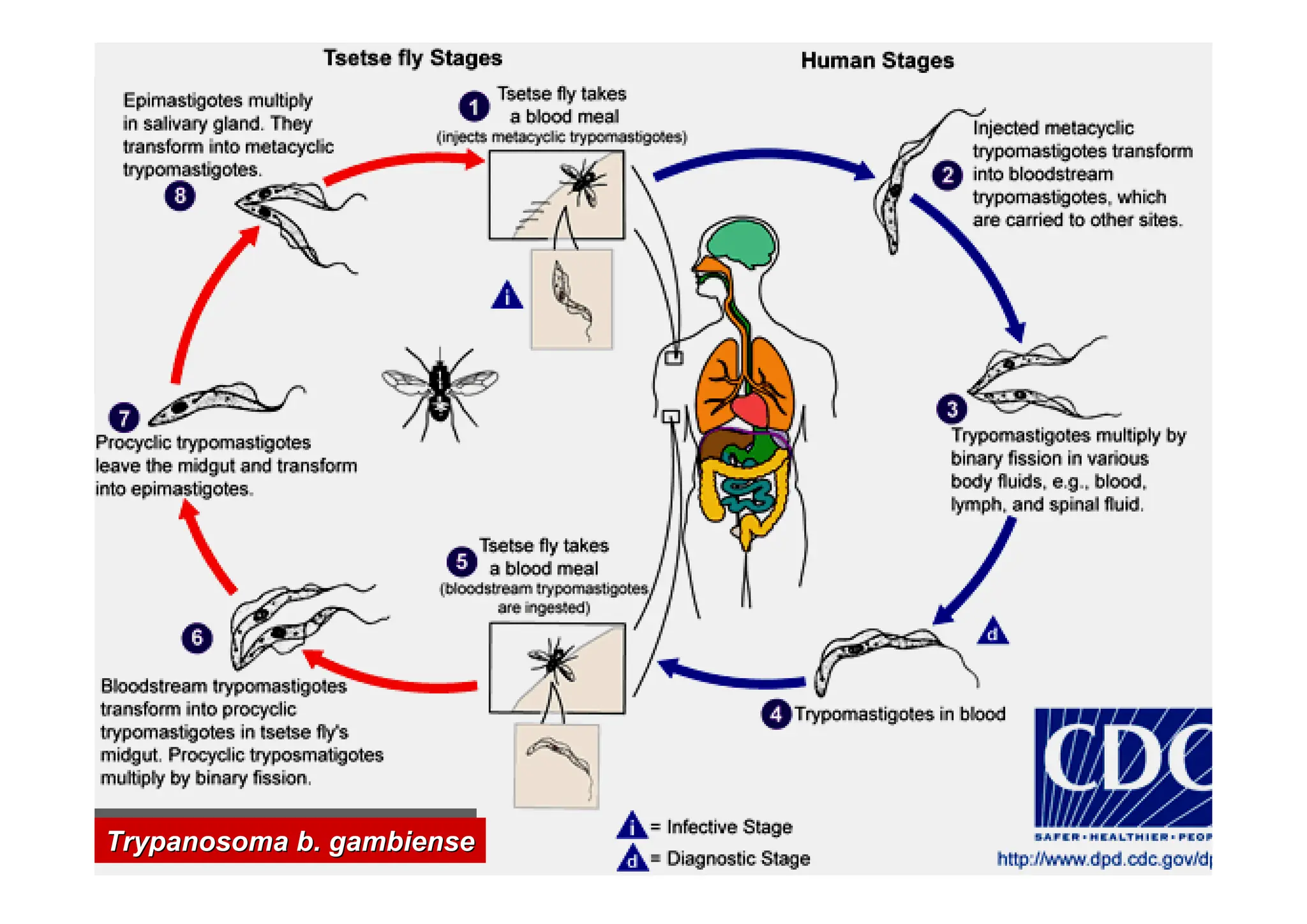
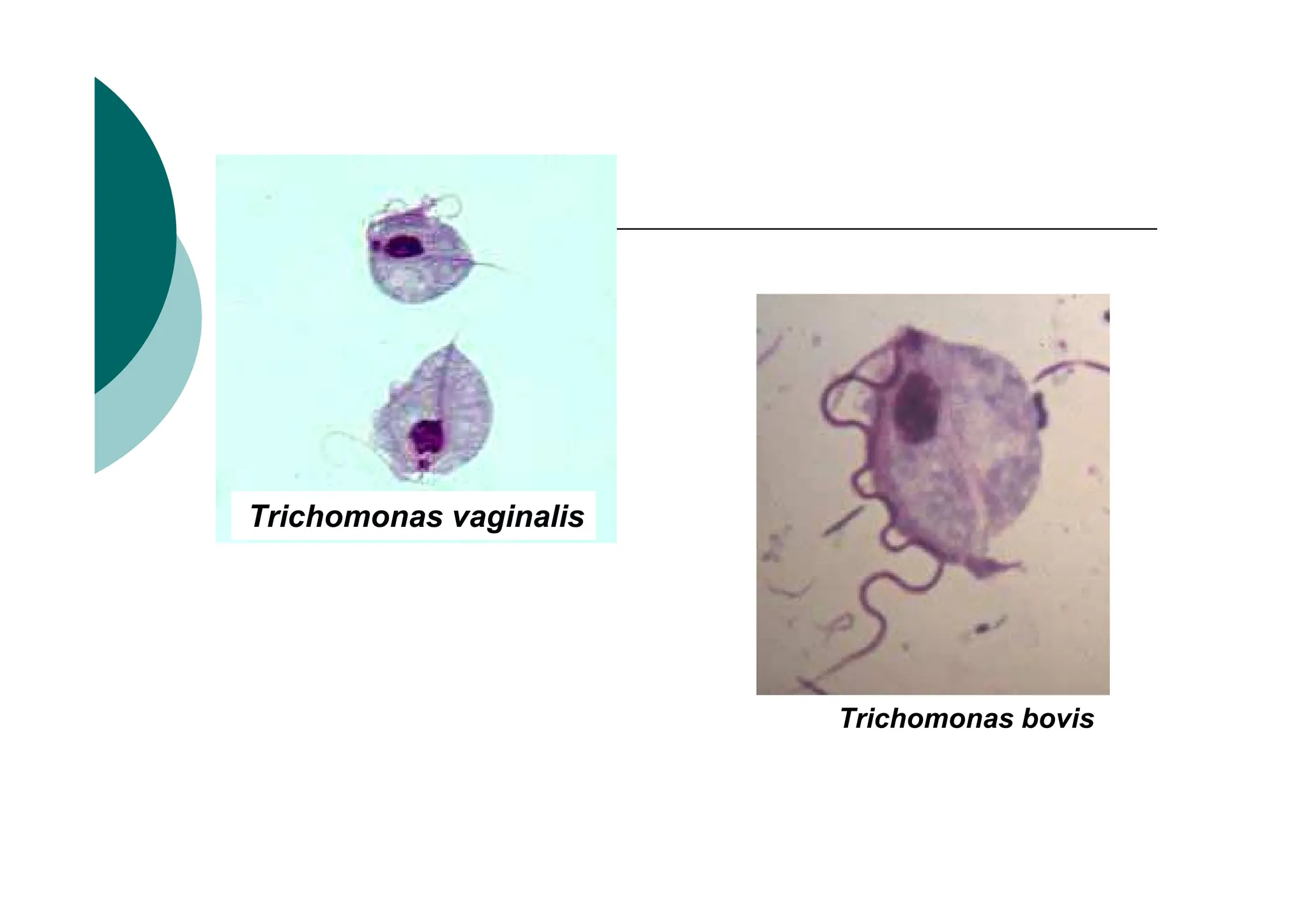
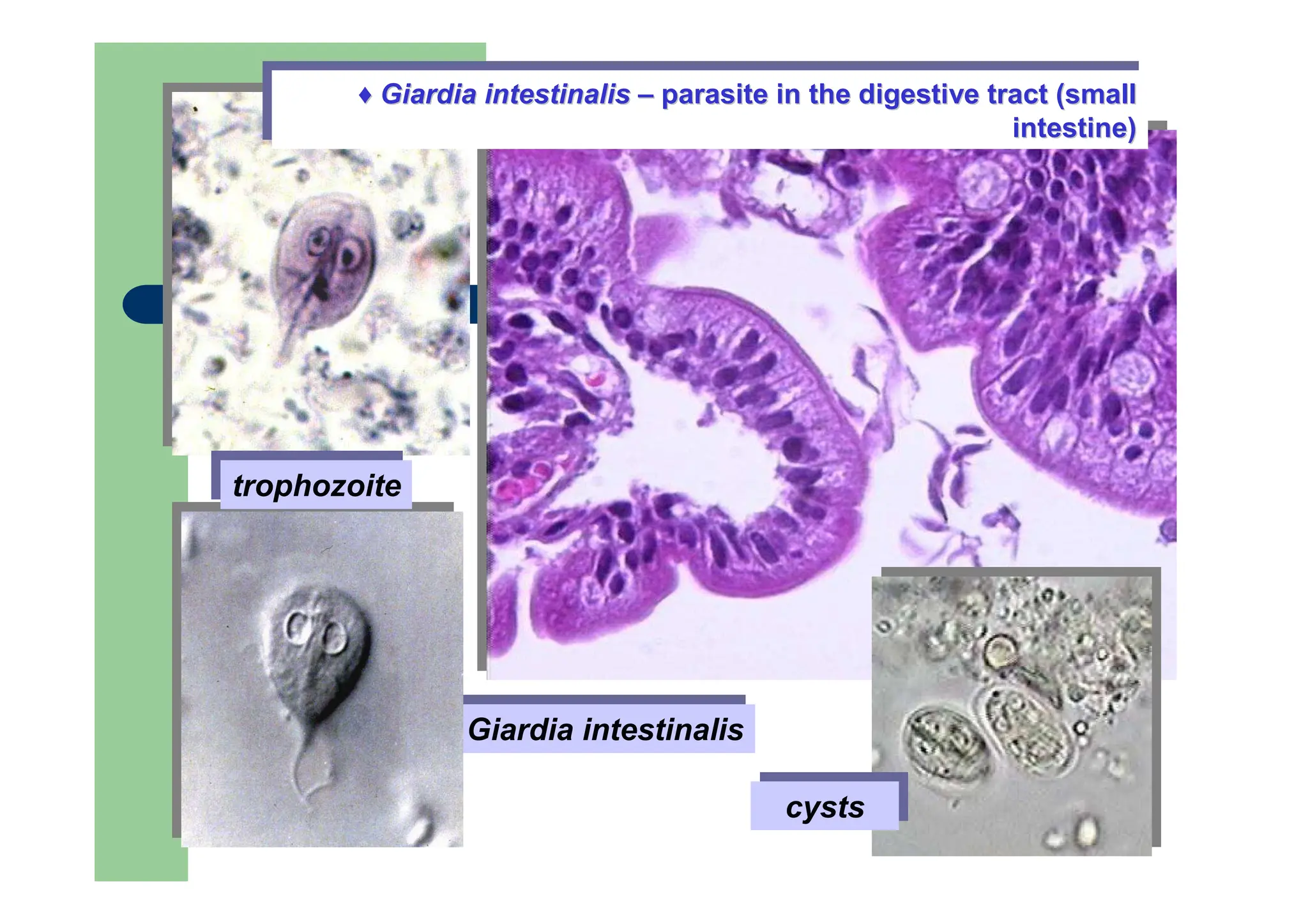
Examples: Trypanosoma spp., Trichomonas spp.,
Giardia intestinalis, Ichthyobodor necator
Trypanosoma – causes African trypanosomiasis or sleeping sickness - a parasitic
disease of humans and other animals. It is caused by protozoa of the species
Trypanosoma brucei
-There are two types that infect humans, Trypanosoma brucei gambiense (T.b.g)
and Trypanosoma brucei rhodesiense (T.b.r.). T.b.g causes over 98% of reported
cases.
-T. brucei brucei causes a related disease in domestic animals (known as nagana).
- both are usually transmitted by the bite of an infected tsetse fly.]
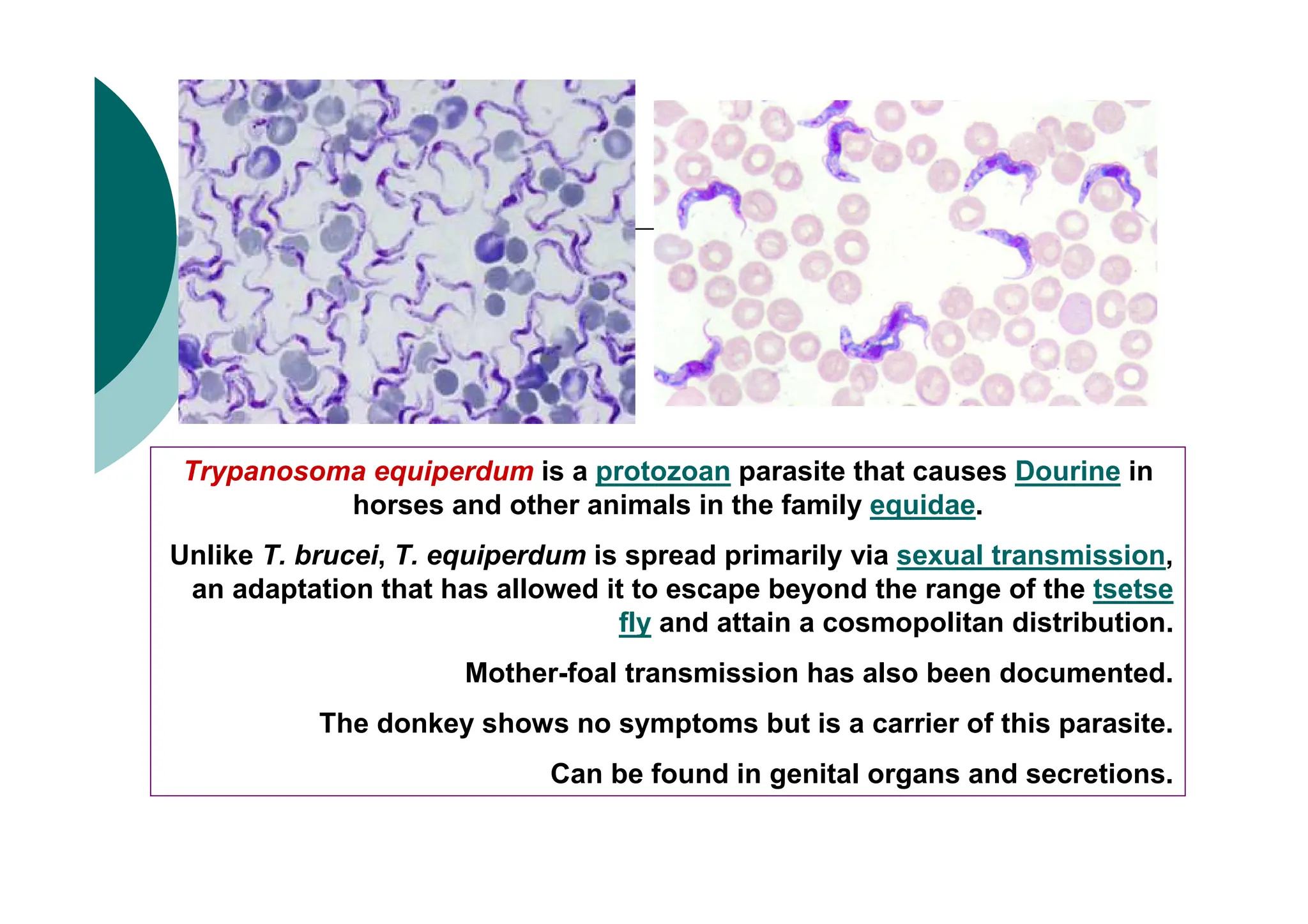
Τrypanosoma equiperdum isa protozoan parasite that causes Dourine in
horses and other animals in the family equidae.
Unlike T. brucei, T. equiperdum is spread primarily via sexual transmission,
an adaptation that has allowed it to escape beyond the range of the tsetse
fly and attain a cosmopolitan distribution.
Mother-foal transmission has also been documented.
Τhe donkey shows no symptoms but is a carrier of this parasite.
Can be found in genital organs and secretions.
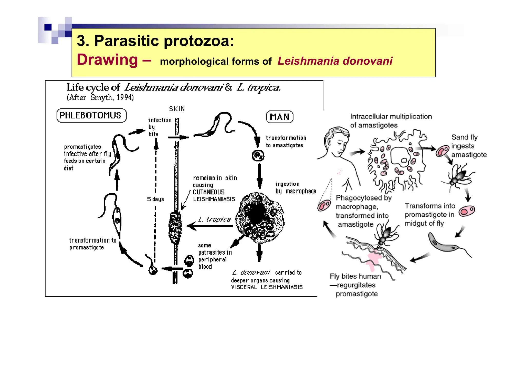
23.
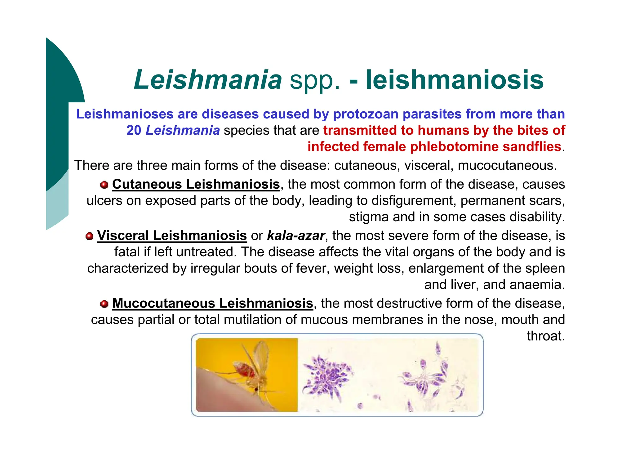
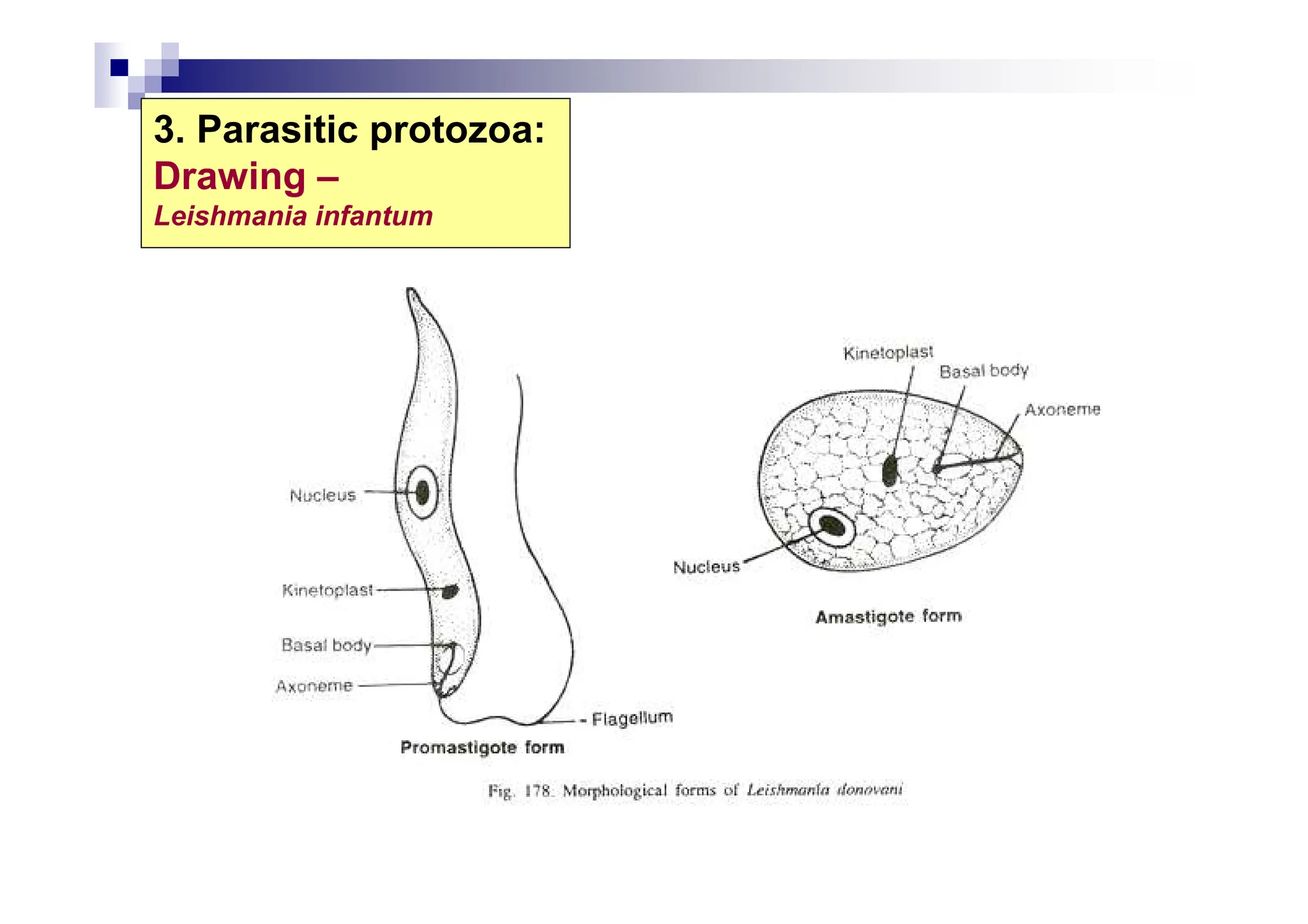
Leishmania spp. -leishmaniosis
Leishmanioses are diseases caused by protozoan parasites from more than
20 Leishmania species that are transmitted to humans by the bites of
infected female phlebotomine sandflies.
There are three main forms of the disease: cutaneous, visceral, mucocutaneous.
Cutaneous Leishmaniosis, the most common form of the disease, causes
ulcers on exposed parts of the body, leading to disfigurement, permanent scars,
stigma and in some cases disability.
Visceral Leishmaniosis or kala-azar, the most severe form of the disease, is
fatal if left untreated. The disease affects the vital organs of the body and is
characterized by irregular bouts of fever, weight loss, enlargement of the spleen
and liver, and anaemia.
Mucocutaneous Leishmaniosis, the most destructive form of the disease,
causes partial or total mutilation of mucous membranes in the nose, mouth and
throat.
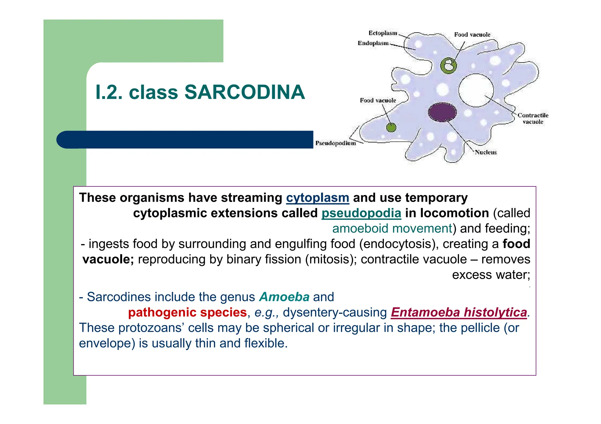
These organisms havestreaming cytoplasm and use temporary
cytoplasmic extensions called pseudopodia in locomotion (called
amoeboid movement) and feeding;
- ingests food by surrounding and engulfing food (endocytosis), creating a food
vacuole; reproducing by binary fission (mitosis); contractile vacuole – removes
excess water;
.
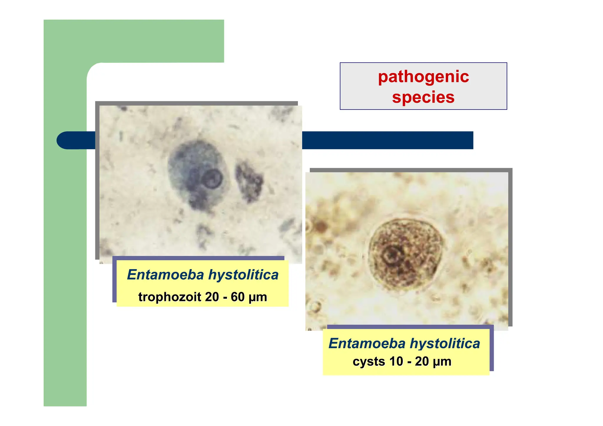
- Sarcodines include the genus Amoeba and
pathogenic species, e.g., dysentery-causing Entamoeba histolytica.
These protozoans’ cells may be spherical or irregular in shape; the pellicle (or
envelope) is usually thin and flexible.
I.2. class SARCODINA
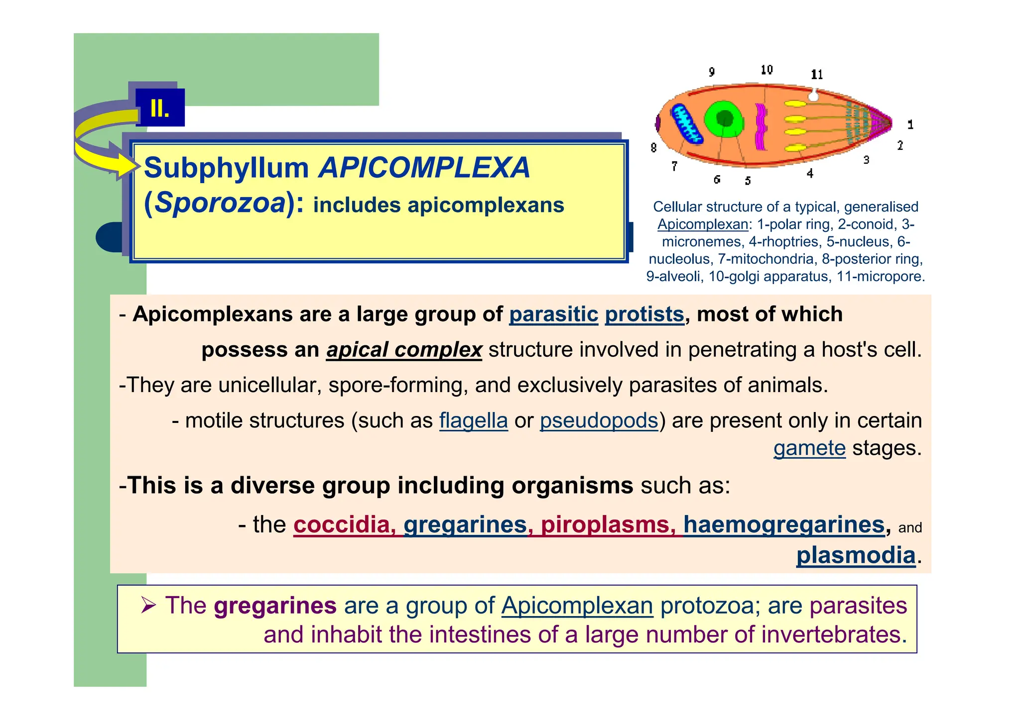
Subphyllum APICOMPLEXA
(Sporozoa): includesapicomplexans
Subphyllum APICOMPLEXA
(Sporozoa): includes apicomplexans
II.
II.
- Apicomplexans are a large group of parasitic protists, most of which
possess an apical complex structure involved in penetrating a host's cell.
-They are unicellular, spore-forming, and exclusively parasites of animals.
- motile structures (such as flagella or pseudopods) are present only in certain
gamete stages.
-This is a diverse group including organisms such as:
- the coccidia, gregarines, piroplasms, haemogregarines, and
plasmodia.
Cellular structure of a typical, generalised
Apicomplexan: 1-polar ring, 2-conoid, 3-
micronemes, 4-rhoptries, 5-nucleus, 6-
nucleolus, 7-mitochondria, 8-posterior ring,
9-alveoli, 10-golgi apparatus, 11-micropore.
The gregarines are a group of Apicomplexan protozoa; are parasites
and inhabit the intestines of a large number of invertebrates.
30.
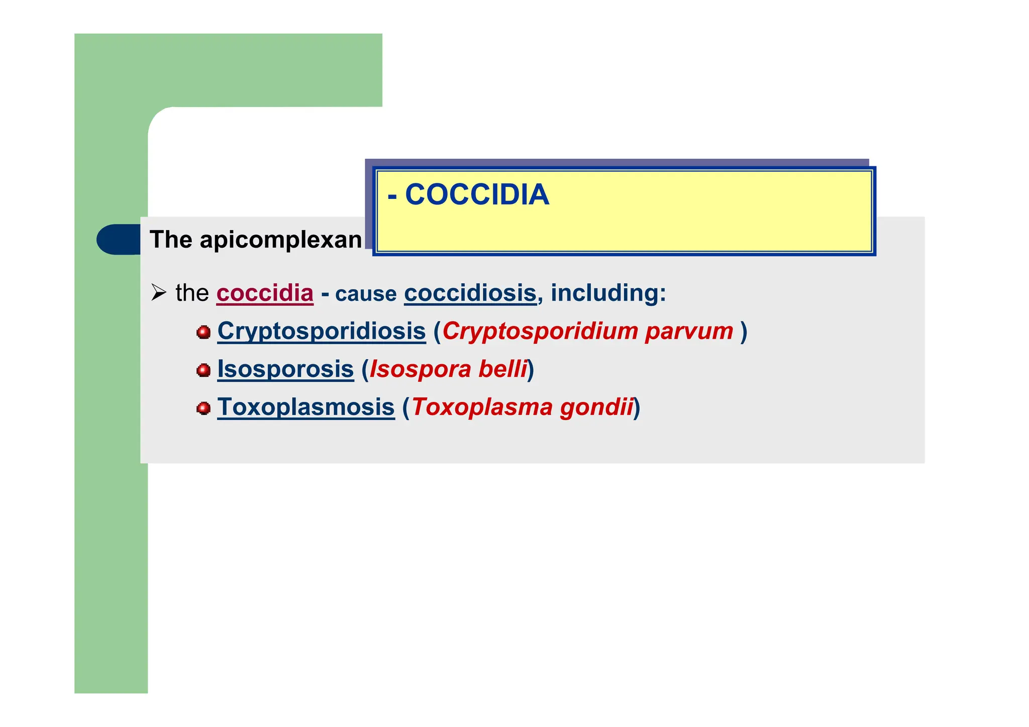
The apicomplexan organismsinclude:
the coccidia - cause coccidiosis, including:
Cryptosporidiosis (Cryptosporidium parvum )
Isosporosis (Isospora belli)
Toxoplasmosis (Toxoplasma gondii)
- COCCIDIA
- COCCIDIA
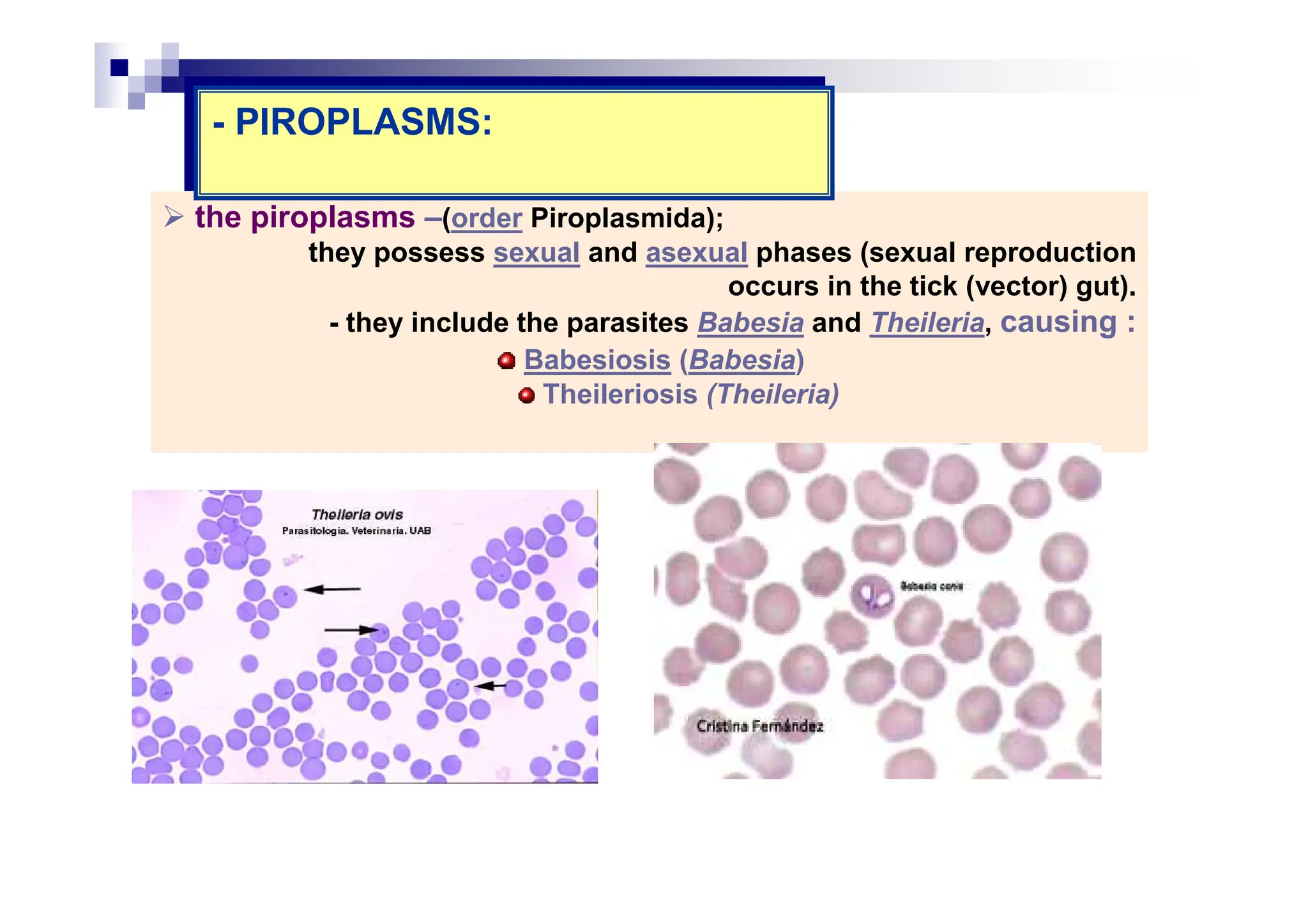
the piroplasms –(orderPiroplasmida);
they possess sexual and asexual phases (sexual reproduction
occurs in the tick (vector) gut).
- they include the parasites Babesia and Theileria, causing :
Babesiosis (Babesia)
Theileriosis (Theileria)
- PIROPLASMS:
- PIROPLASMS:
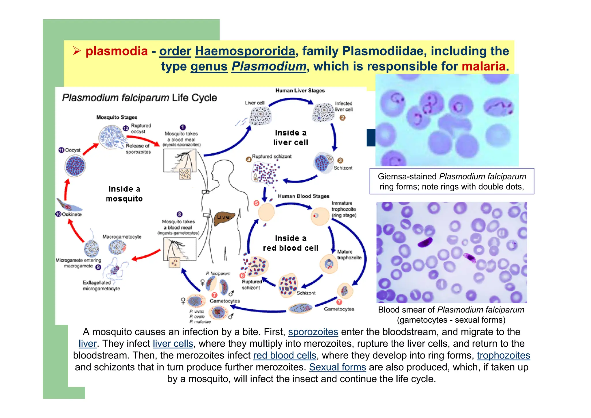
plasmodia - orderHaemospororida, family Plasmodiidae, including the
type genus Plasmodium, which is responsible for malaria.
A mosquito causes an infection by a bite. First, sporozoites enter the bloodstream, and migrate to the
liver. They infect liver cells, where they multiply into merozoites, rupture the liver cells, and return to the
bloodstream. Then, the merozoites infect red blood cells, where they develop into ring forms, trophozoites
and schizonts that in turn produce further merozoites. Sexual forms are also produced, which, if taken up
by a mosquito, will infect the insect and continue the life cycle.
Blood smear of Plasmodium falciparum
(gametocytes - sexual forms)
Giemsa-stained Plasmodium falciparum
ring forms; note rings with double dots,
35.
Subphylum
CILIATA (Ciliophora):
Subphylum
CILIATA (Ciliophora):
III.
III.
-The ciliates are a group of protozoans characterized by the presence of
hair-like organelles called cilia,
- in general, are larger than any other protozoa, range in length from 10 µm
to 3 mm);
- are the most structurally complex of all protozoans, exhibiting a wide
range of specializations.
- multinucleate, possessing one macronucleus (with metabolic role) and one
micronucleus (genetic role).
- nutrition heterotrophic; most of them, posses a cytostome (mouth)- a simple
opening /or connected to a gullet or ciliated groove;
-- contractile valuoles typically present;
- most species free living, but many commensal, some parasitic.
- The ciliates are a group of protozoans characterized by the presence of
hair-like organelles called cilia,
- in general, are larger than any other protozoa, range in length from 10 µm
to 3 mm);
- are the most structurally complex of all protozoans, exhibiting a wide
range of specializations.
- multinucleate, possessing one macronucleus (with metabolic role) and one
micronucleus (genetic role).
- nutrition heterotrophic; most of them, posses a cytostome (mouth)- a simple
opening /or connected to a gullet or ciliated groove;
-- contractile valuoles typically present;
- most species free living, but many commensal, some parasitic.
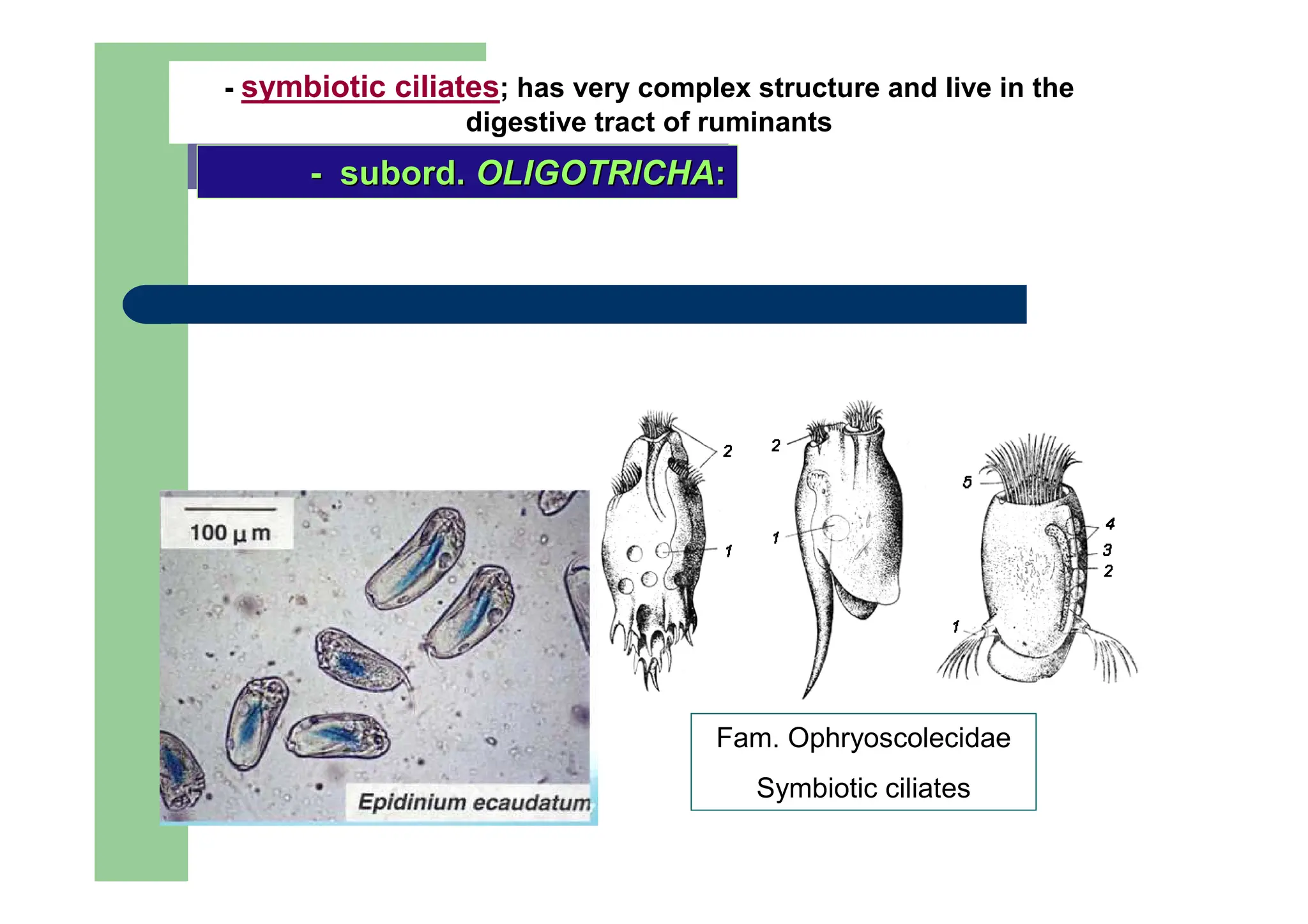
- subord. OLIGOTRICHA:
-
-subord.
subord. OLIGOTRICHA
OLIGOTRICHA:
:
- symbiotic ciliates; has very complex structure and live in the
digestive tract of ruminants
Fam. Ophryoscolecidae
Symbiotic ciliates
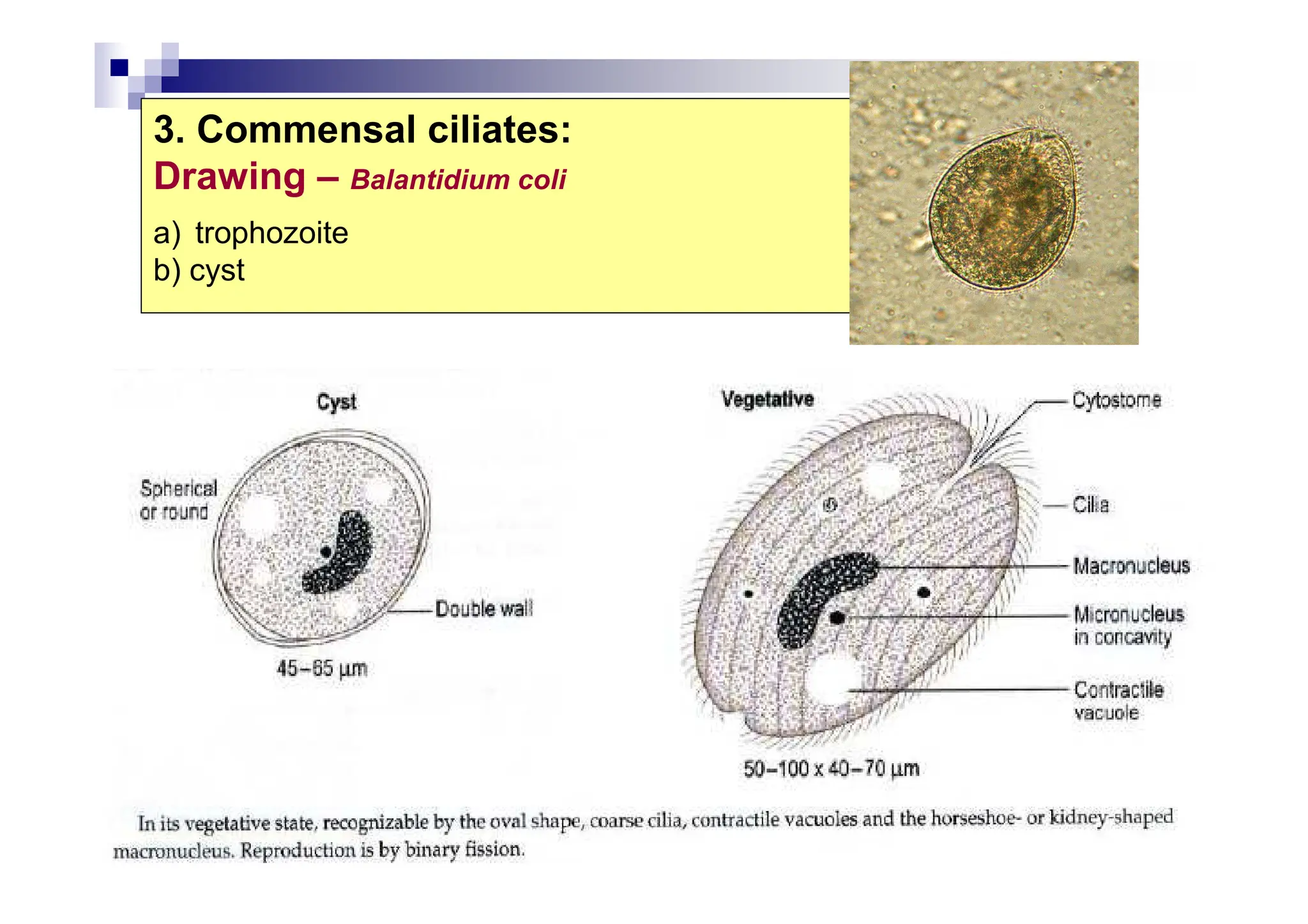
38.
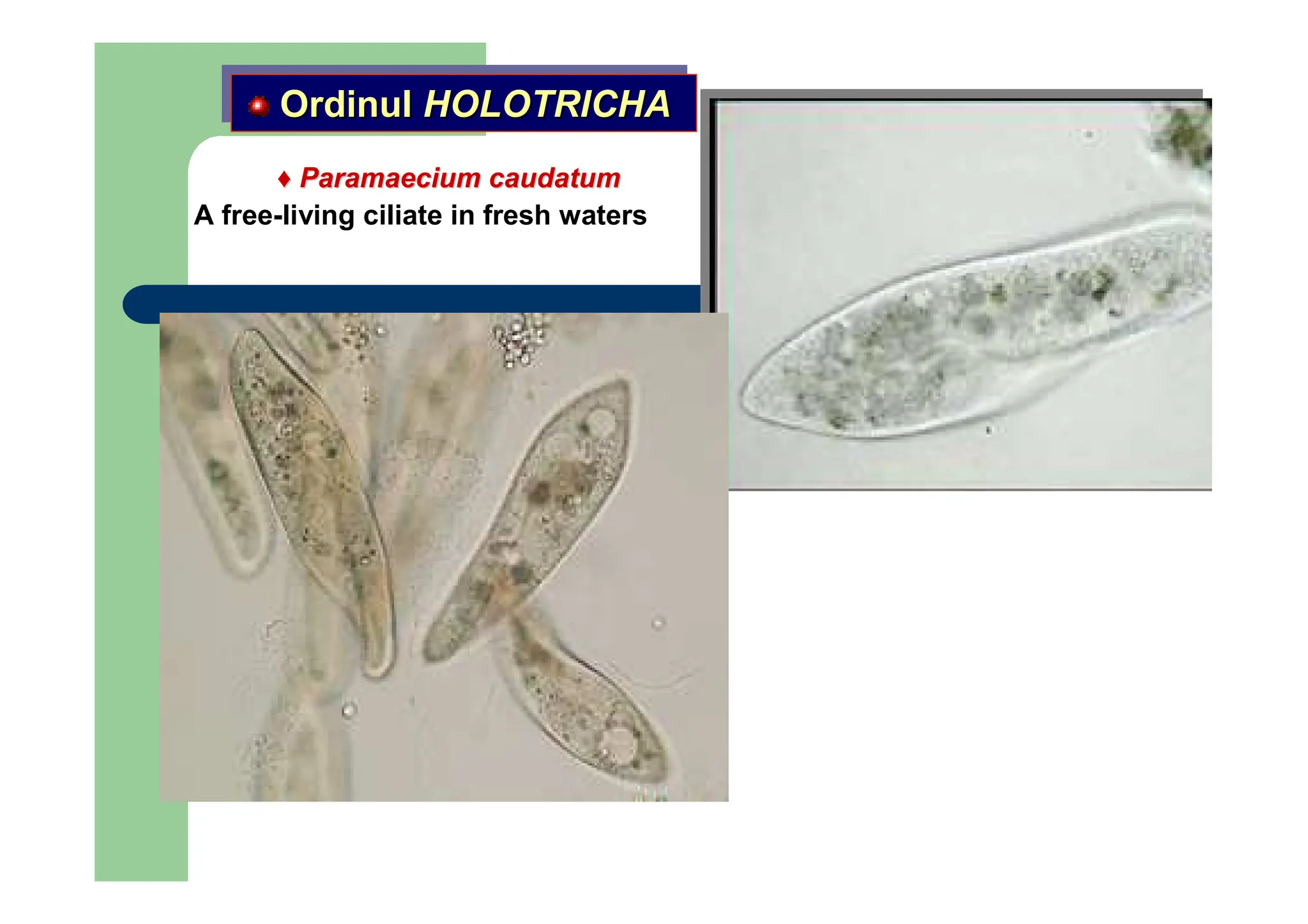
Balantidium coli
- trophozoite-
-Lives as commensal in the large intestine of humans, pigs,
rats . Usually is not pathogenic, but in some cases invade the
intestinal lining (becomes parasites) and cause disease -
disentery.
Ordinul SPIROTRICHA
Ordinul
Ordinul SPIROTRICHA
SPIROTRICHA
39.
Subphylum MICROSPORA
(microsporidia):
spore-forming unicellularparasites
Subphylum MICROSPORA
(mi
micr
crosporidi
osporidia
a):
spore-forming unicellular parasites
IV.
IV.
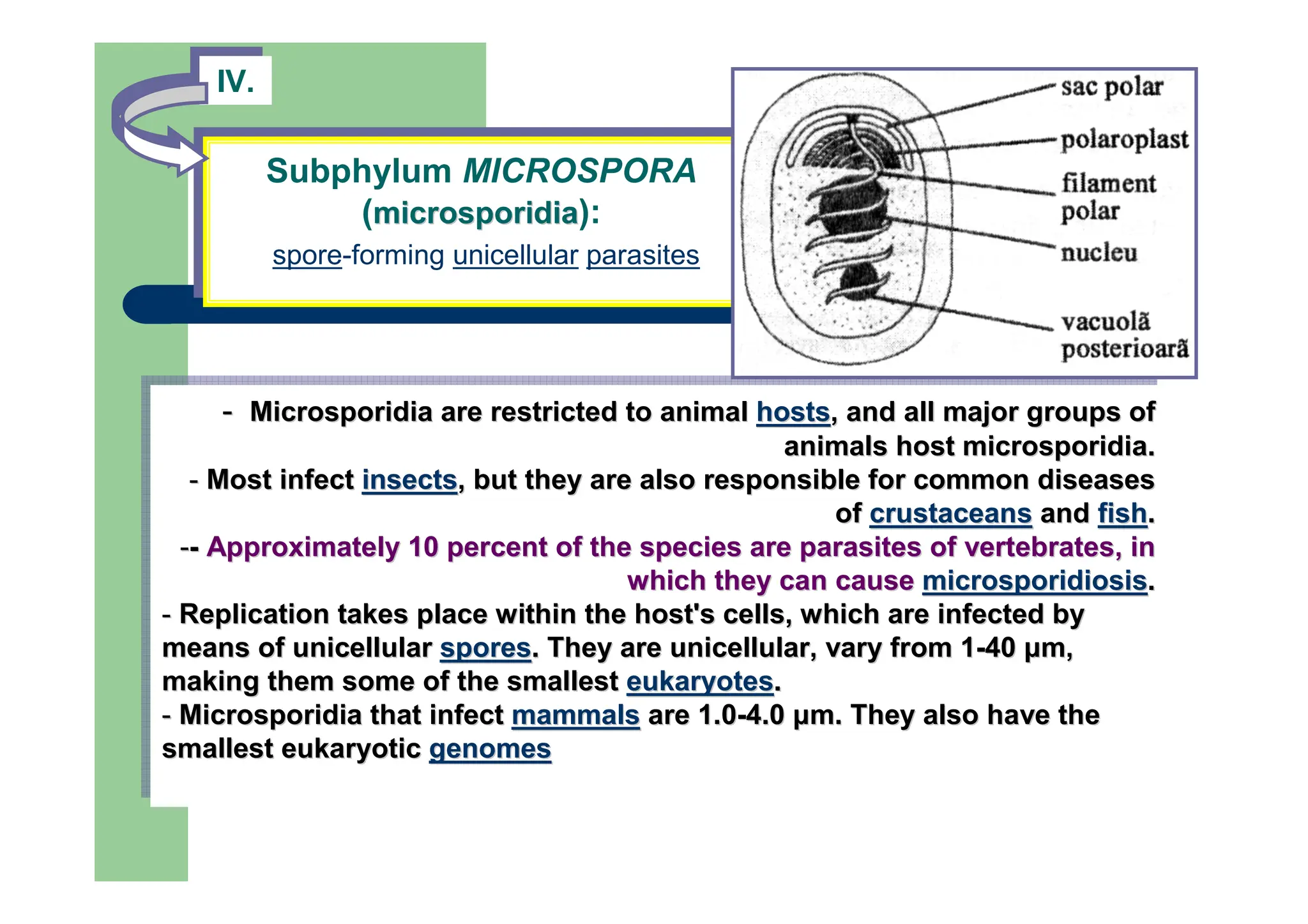
- Microsporidia are restricted to animal hosts, and all major groups of
animals host microsporidia.
- Most infect insects, but they are also responsible for common diseases
of crustaceans and fish.
-- Approximately 10 percent of the species are parasites of vertebrates, in
which they can cause microsporidiosis.
- Replication takes place within the host's cells, which are infected by
means of unicellular spores. They are unicellular, vary from 1-40 µm,
making them some of the smallest eukaryotes.
- Microsporidia that infect mammals are 1.0-4.0 µm. They also have the
smallest eukaryotic genomes
-
- Microsporidia
Microsporidia are restricted to animal
are restricted to animal hosts
hosts, and all major groups of
, and all major groups of
animals host
animals host microsporidia
microsporidia.
.
-
- Most infect
Most infect insects
insects, but they are also responsible for common diseases
, but they are also responsible for common diseases
of
of crustaceans
crustaceans and
and fish
fish.
.
-
--
- Approximately 10 percent of the species are parasites of vertebr
Approximately 10 percent of the species are parasites of vertebrates, in
ates, in
which they can cause
which they can cause microsporidiosis
microsporidiosis.
.
-
- Replication takes place within the host's cells, which are infe
Replication takes place within the host's cells, which are infected by
cted by
means of unicellular
means of unicellular spores
spores. They are unicellular, vary from 1
. They are unicellular, vary from 1-
-40
40 µ
µm
m,
,
making them some of the smallest
making them some of the smallest eukaryotes
eukaryotes.
.
-
- Microsporidia
Microsporidia that infect
that infect mammals
mammals are 1.0
are 1.0-
-4.0
4.0 µ
µm
m. They also have the
. They also have the
smallest eukaryotic
smallest eukaryotic genomes
genomes
,
,
40.
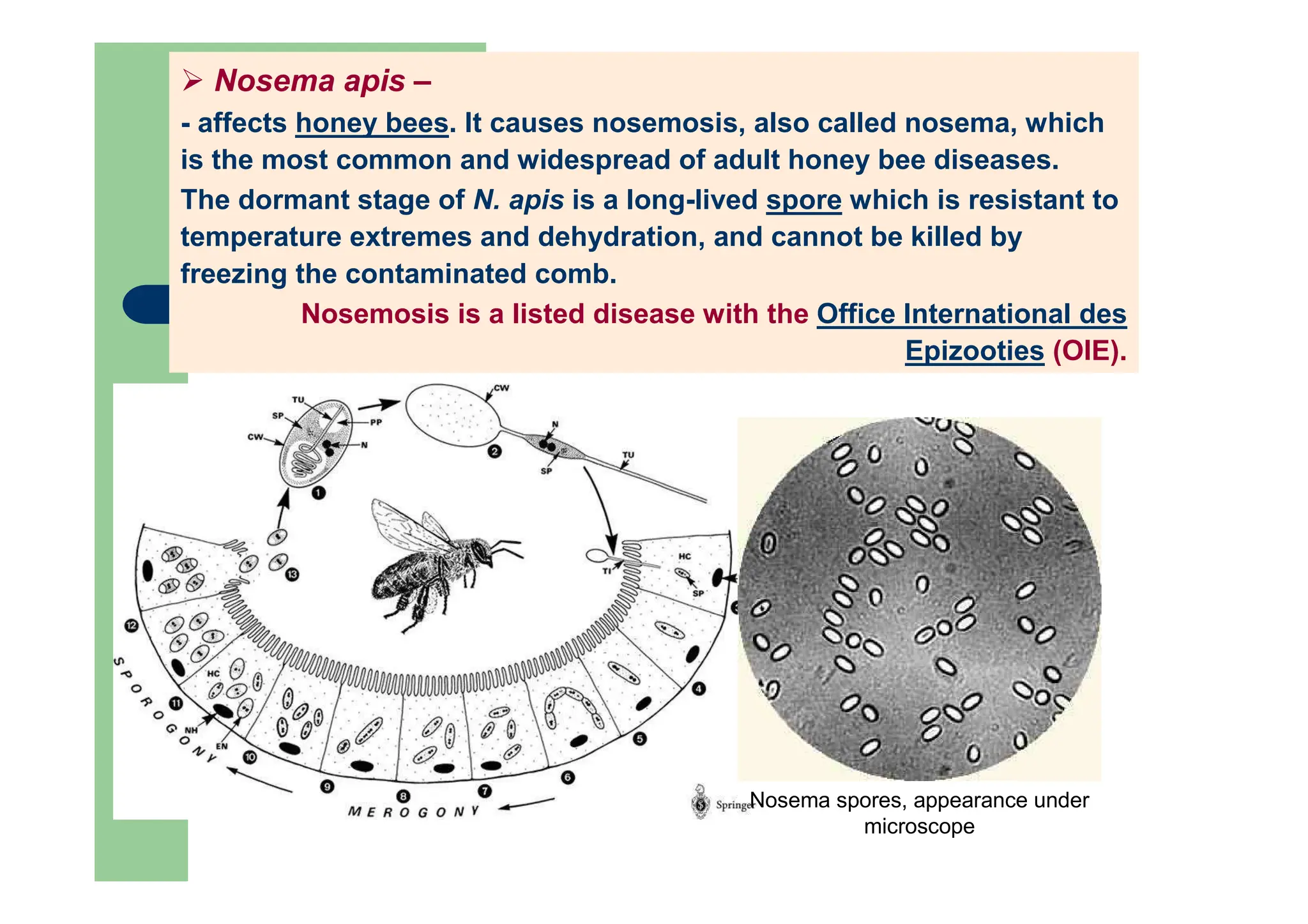
Nosema apis –
-affects honey bees. It causes nosemosis, also called nosema, which
is the most common and widespread of adult honey bee diseases.
The dormant stage of N. apis is a long-lived spore which is resistant to
temperature extremes and dehydration, and cannot be killed by
freezing the contaminated comb.
Nosemosis is a listed disease with the Office International des
Epizooties (OIE).
Nosema spores, appearance under
microscope
41.
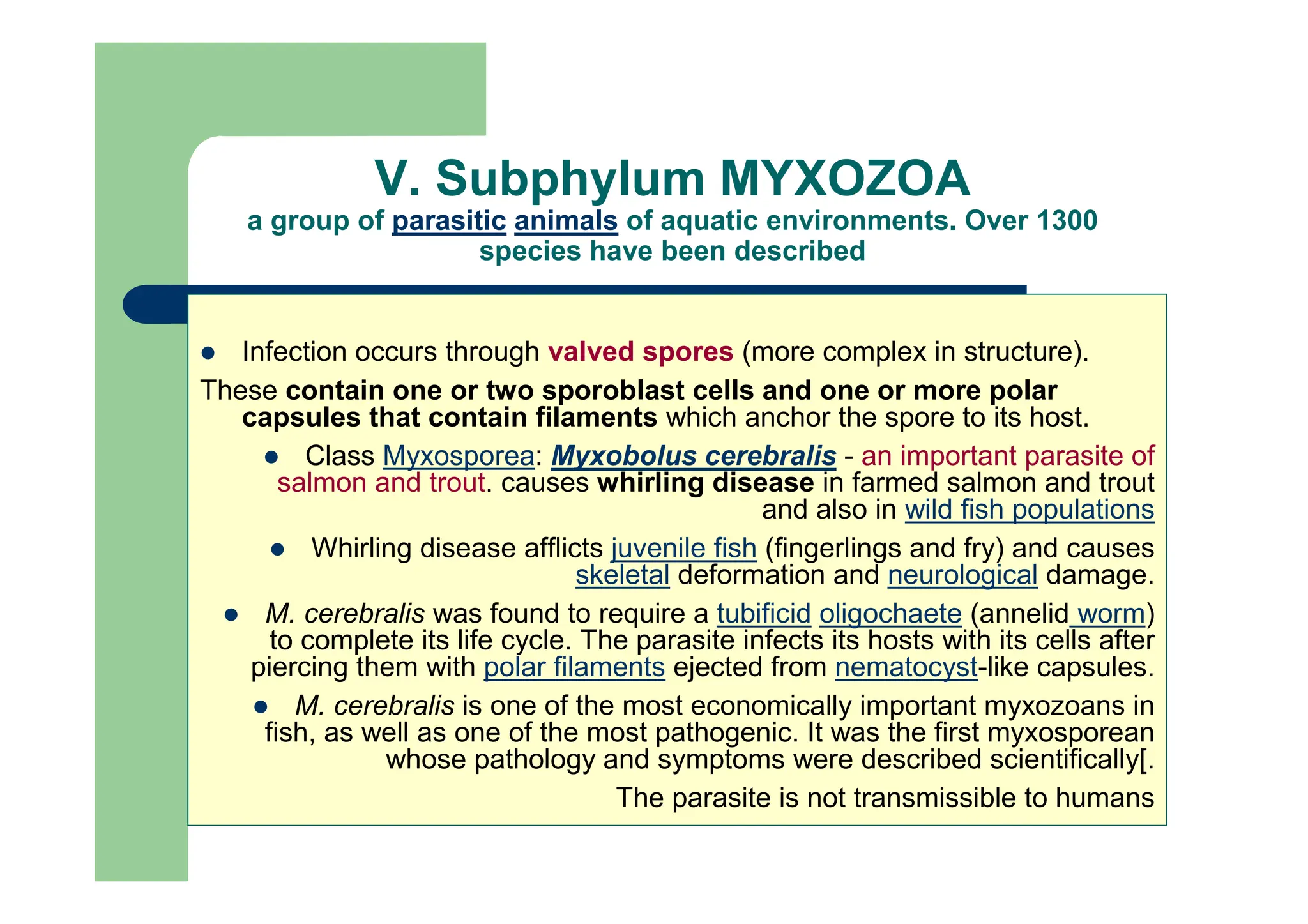
V. Subphylum MYXOZOA
agroup of parasitic animals of aquatic environments. Over 1300
species have been described
Infection occurs through valved spores (more complex in structure).
These contain one or two sporoblast cells and one or more polar
capsules that contain filaments which anchor the spore to its host.
Class Myxosporea: Myxobolus cerebralis - an important parasite of
salmon and trout. causes whirling disease in farmed salmon and trout
and also in wild fish populations
Whirling disease afflicts juvenile fish (fingerlings and fry) and causes
skeletal deformation and neurological damage.
M. cerebralis was found to require a tubificid oligochaete (annelid worm)
to complete its life cycle. The parasite infects its hosts with its cells after
piercing them with polar filaments ejected from nematocyst-like capsules.
M. cerebralis is one of the most economically important myxozoans in
fish, as well as one of the most pathogenic. It was the first myxosporean
whose pathology and symptoms were described scientifically[.
The parasite is not transmissible to humans
42.
I. General characteristicsof Protozoan
1. Unicellular eukaryotes, some colonial, and some with multicellular stages in
their life cycles
2. Mostly microscopic.
3. All symmetries represented in the group; shape variable or constant
(oval, spheric, or other)
4. No organ or tissues, but specialized organelles are found;
nucleus single or multiple.
5. Free-living, mutualism, commensalism, parasitism all represented I the groups.
6. Locomotion by pseudopodia, flagella, cillia, and direct cell movements;
some sessile.
7. Some provided with a simple endoskeleton or exoskeleton, but most are naked.
8. Nutrition of all types: autotrophic, heterotrophic, saprozoic (using nutrients
dissolved in the surrounding medium)
9. Reproduction asexually by fission, budding, and cysts and sexually, by
conjugation or syngamy (union of a male and female gametes to form a zygote).
9. Aquatic or terrestrial habitat; free-living, symbiotic, commensal,or parasitic
mode of life.
Revision / Key Notes:
Revision / Key Notes:
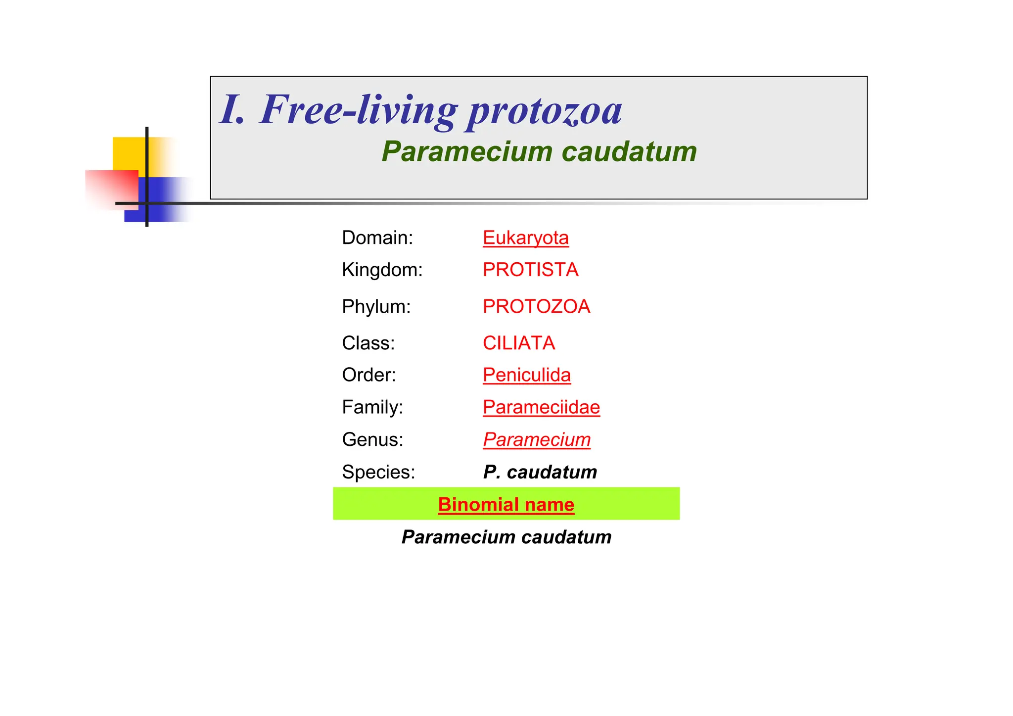
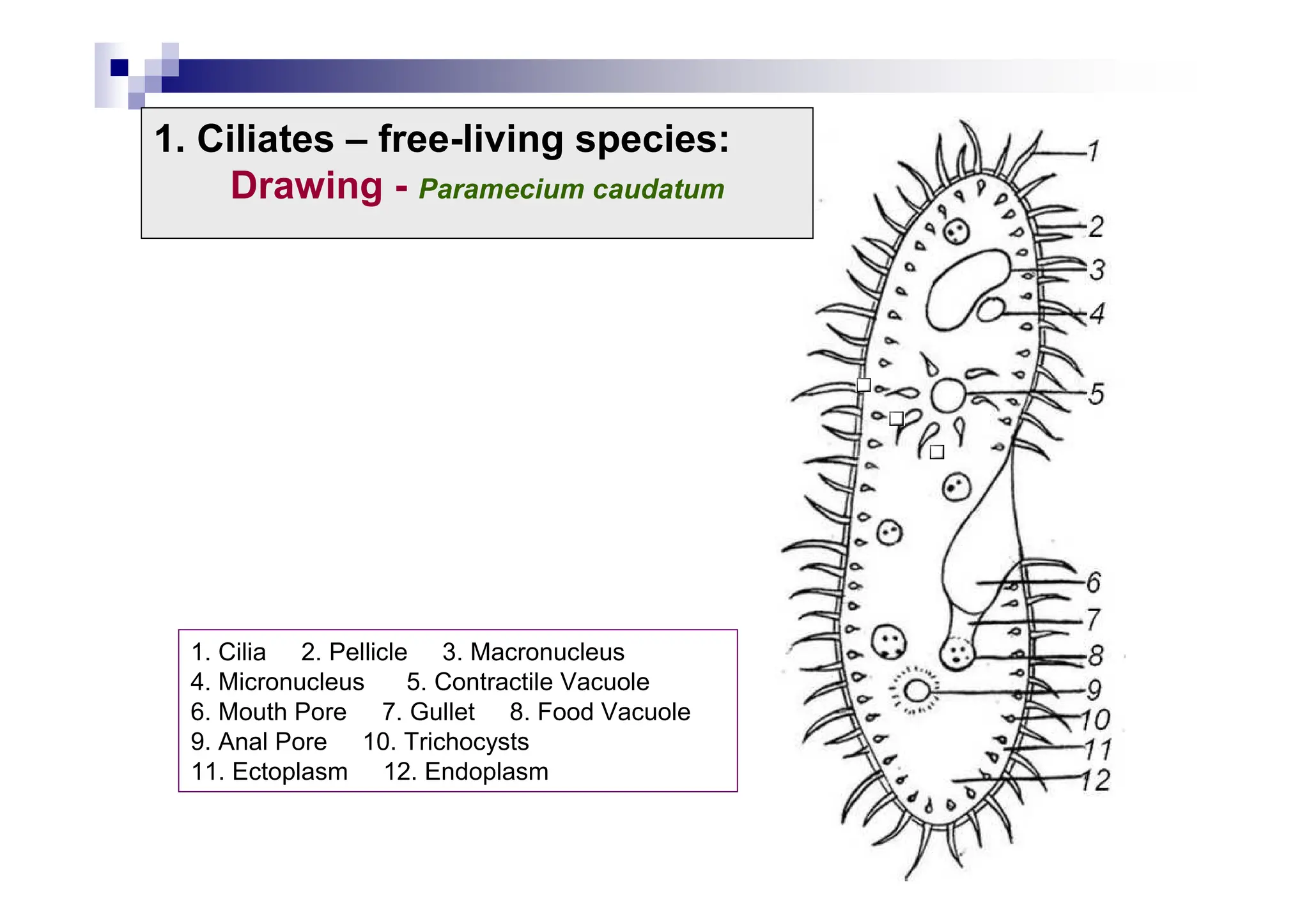
I. Free-living protozoa
Parameciumcaudatum
Domain: Eukaryota
Kingdom: PROTISTA
Phylum: PROTOZOA
Class: CILIATA
Order: Peniculida
Family: Parameciidae
Genus: Paramecium
Species: P. caudatum
Binomial name
Paramecium caudatum
45.
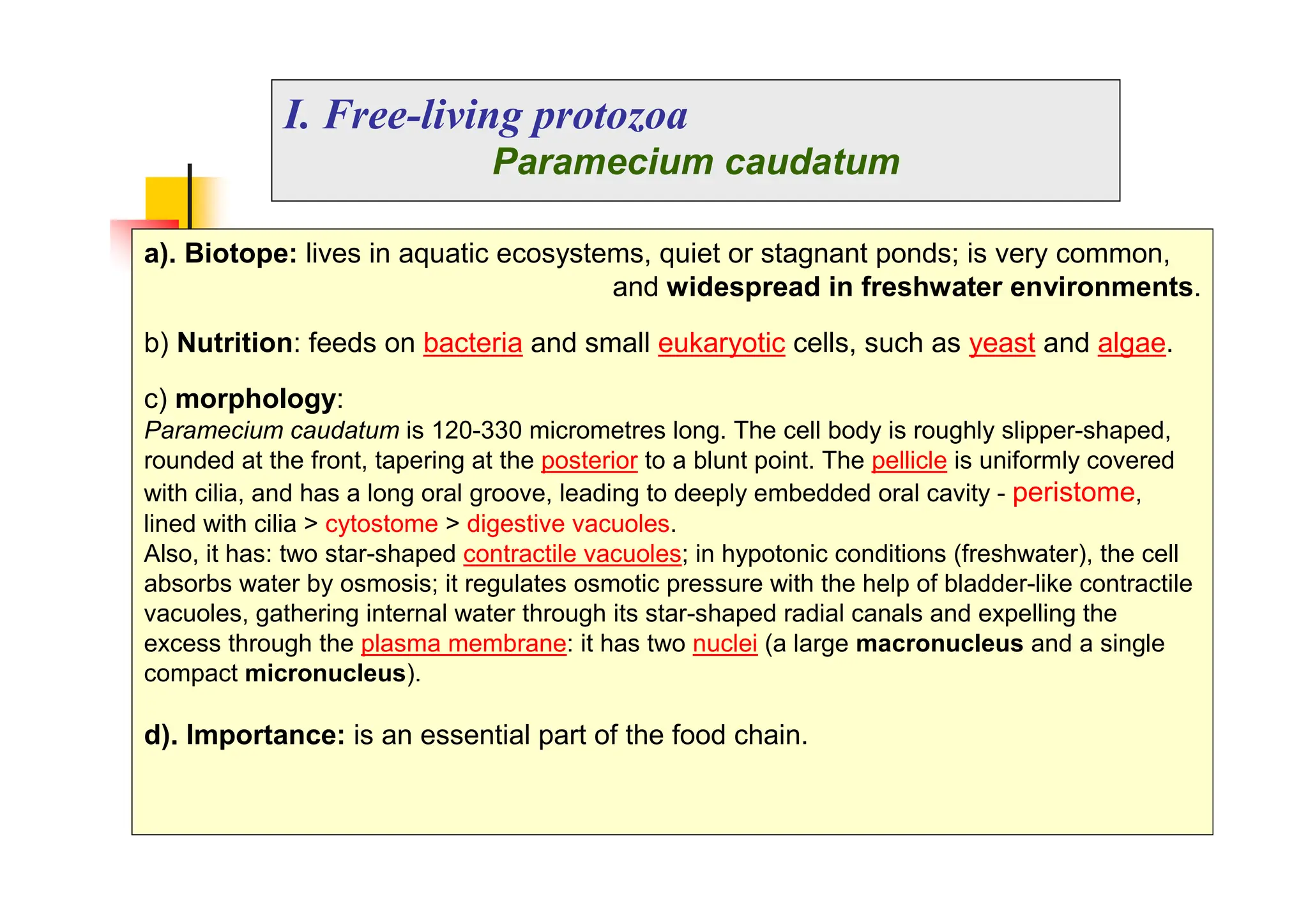
I. Free-living protozoa
Parameciumcaudatum
a). Biotope: lives in aquatic ecosystems, quiet or stagnant ponds; is very common,
and widespread in freshwater environments.
b) Nutrition: feeds on bacteria and small eukaryotic cells, such as yeast and algae.
c) morphology:
Paramecium caudatum is 120-330 micrometres long. The cell body is roughly slipper-shaped,
rounded at the front, tapering at the posterior to a blunt point. The pellicle is uniformly covered
with cilia, and has a long oral groove, leading to deeply embedded oral cavity - peristome,
lined with cilia > cytostome > digestive vacuoles.
Also, it has: two star-shaped contractile vacuoles; in hypotonic conditions (freshwater), the cell
absorbs water by osmosis; it regulates osmotic pressure with the help of bladder-like contractile
vacuoles, gathering internal water through its star-shaped radial canals and expelling the
excess through the plasma membrane: it has two nuclei (a large macronucleus and a single
compact micronucleus).
d). Importance: is an essential part of the food chain.
46.
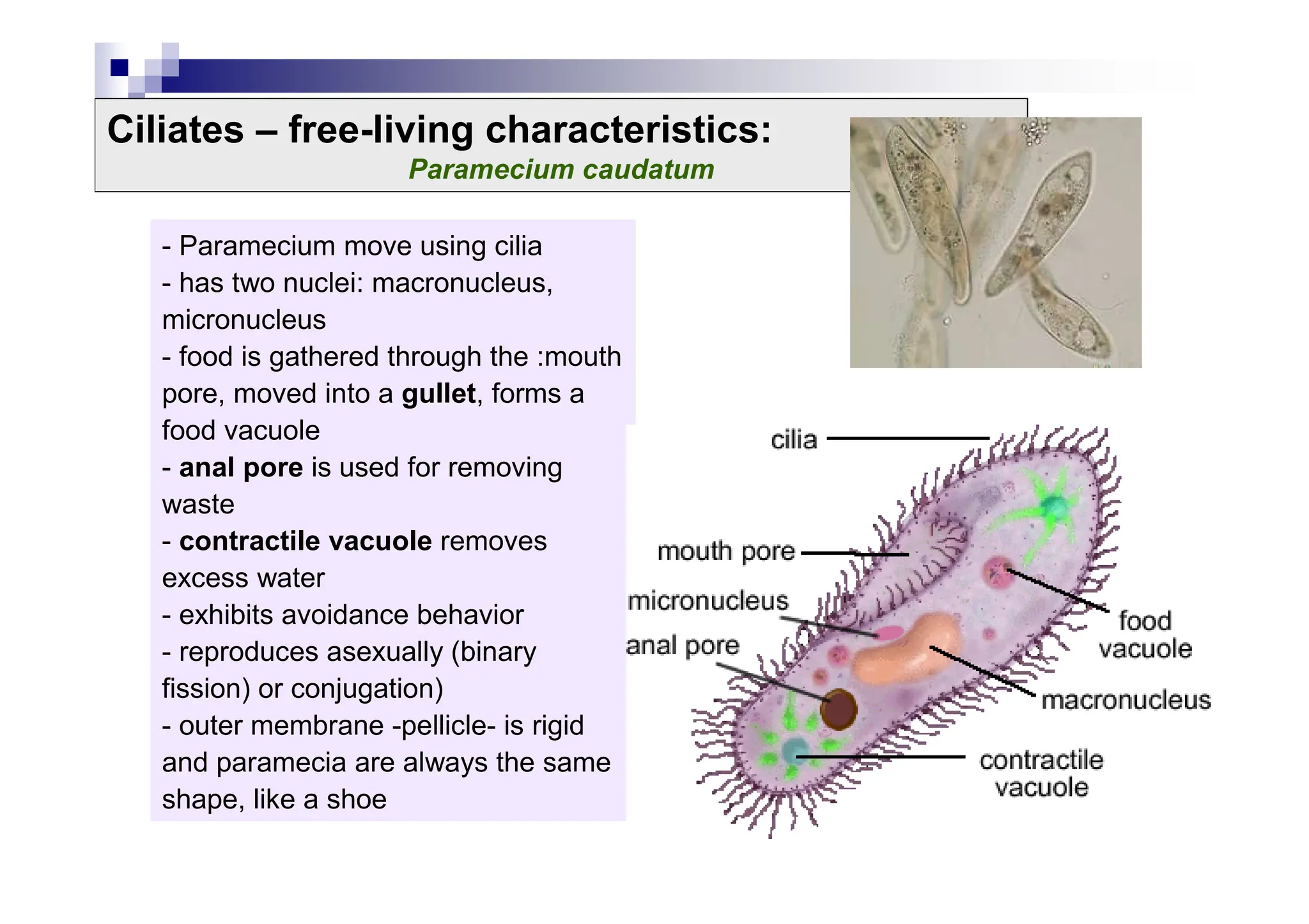
Ciliates – free-livingcharacteristics:
Paramecium caudatum
- Paramecium move using cilia
- has two nuclei: macronucleus,
micronucleus
- food is gathered through the :mouth
pore, moved into a gullet, forms a
food vacuole
- anal pore is used for removing
waste
- contractile vacuole removes
excess water
- exhibits avoidance behavior
- reproduces asexually (binary
fission) or conjugation)
- outer membrane -pellicle- is rigid
and paramecia are always the same
shape, like a shoe
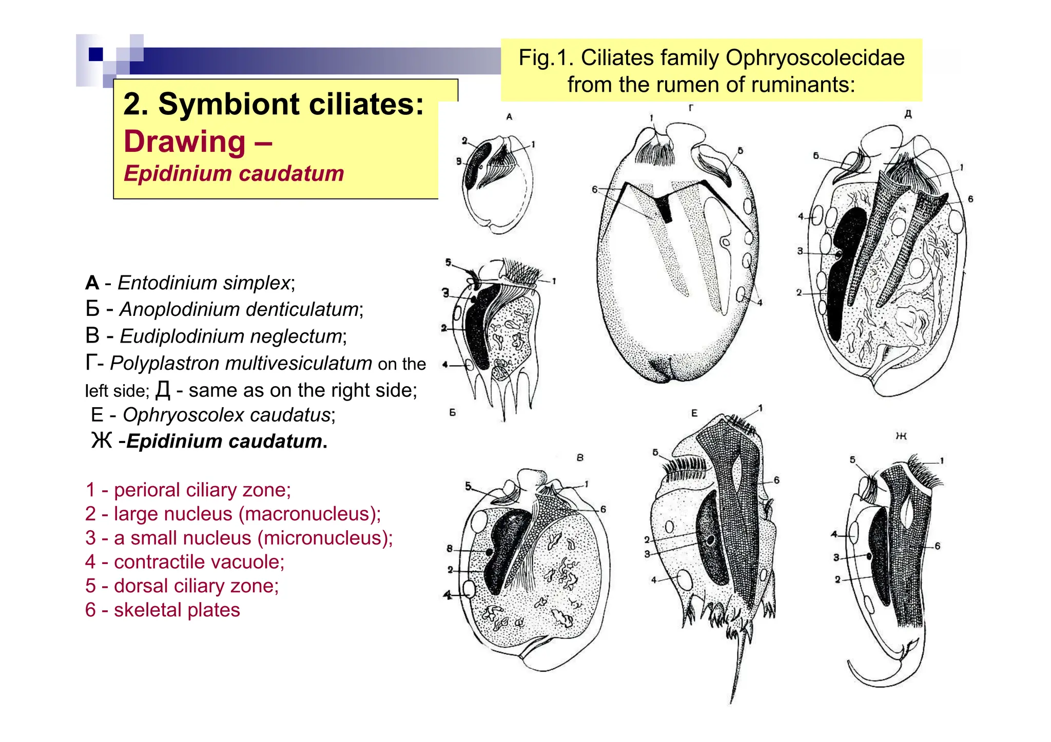
2. Symbiont ciliates:
Drawing–
Epidinium caudatum
A - Entodinium simplex;
Б - Anoplodinium denticulatum;
В - Eudiplodinium neglectum;
Г- Polyplastron multivesiculatum on the
left side; Д - same as on the right side;
E - Ophryoscolex caudatus;
Ж -Epidinium caudatum.
1 - perioral ciliary zone;
2 - large nucleus (macronucleus);
3 - a small nucleus (micronucleus);
4 - contractile vacuole;
5 - dorsal ciliary zone;
6 - skeletal plates
Fig.1. Ciliates family Ophryoscolecidae
from the rumen of ruminants:
![Protozoa are a diverse group of unicellular eukaryotic organisms,
- usually single-celled and heterotrophic eukaryotes containing non-
filamentous structures that belong to any of the major lineages of protists.
- They are restricted to moist or aquatic habitats:
-i.e., they are obligate aquatic organisms;
- many protozoan species are symbionts,
- some are parasites,
- and some are predators of bacteria and algae.
There are an estimated 30,000 protozoan species]
Protozoa are a diverse group of unicellular eukaryotic organisms,
- usually single-celled and heterotrophic eukaryotes containing non-
filamentous structures that belong to any of the major lineages of protists.
- They are restricted to moist or aquatic habitats:
-i.e., they are obligate aquatic organisms;
- many protozoan species are symbionts,
- some are parasites,
- and some are predators of bacteria and algae.
There are an estimated 30,000 protozoan species]
Phylum Protozoa Greek protos = first, zoon = animal)
I. Characteristics of protozoa](https://image.slidesharecdn.com/2-251114150927-108b2121/75/2-Course-_Protozoa_Engl-1-pdf-english-section-3-2048.jpg)
![Reproduction
Asexual reproduction:
Binary fission (equal binary fission): in which a cell divides into two
daughter cells (after the chromosomes have been duplicated and distributed
between them). This asexual mode of reproduction leads to rapid population
growth; It may be:
longitudinal
transverse
Internal budding:
Endodiogeny: a new organism develops from an outgrowth or bud due
to cell division at one particular site.
Endopoliogeny: many organisms..
Schizogony (merogony): multiple fission manifested either as:
merogony - results in merozoites: which are multiple daughter cells, that
originate within the same cell membrane:
trophozoite grows to a large size while the nucleus divides repeatedly.
This structure is called a meront (schizont) and, when mature, each
nucleus has acquired a portion of the cytoplasm so that the schizont
is filled with a large number of elongated separate organisms called
merozoites. The meront eventually ruptures, liberating the individual
merozoites.
sporogony results in sporozoites
[Sporogony follows sexual reproduction].
Protozoa: biology](https://image.slidesharecdn.com/2-251114150927-108b2121/75/2-Course-_Protozoa_Engl-1-pdf-english-section-10-2048.jpg)

![Reproduction
Both, asexual and sexual reproduction, by alternative
phases [in Apicomplexa protozoa]: the life cycle consists of:
Schizogony (asexual phase):
Gametogony (sexual phase)
Microgamonts – producing microgametes
Macrogamonts – producing macrogamete.
fecundation - syngamy – results in zygote – egg-cell.
Sporogony – results in oocysts (with sporozoites)
Example: Eimeria (Coccidia) sporulated oocysts have 4
sporoblasts, each with two sporozoites.
Example>
in Eimeria, both asexual and sexual phases occur in the same host
followed by a free-living phase – sporogony (in the environment,
outside of the host).
in others, such as Plasmodium / Babesia, the asexual phase occurs
in the vertebrate host and the sexual phase in the arthropod vector.
Protozoa: biology](https://image.slidesharecdn.com/2-251114150927-108b2121/75/2-Course-_Protozoa_Engl-1-pdf-english-section-12-2048.jpg)
![I.1. Class Mastigophora
• group of Zooflagellata (Zoomastigina):
- protozoa with two or more flagella; some have pseudopodia;
- no color, heterotrophic (holozoice or saprozoice);
- some are free (eg coanoflagelatele); however, the majority are parasitic.
Examples: Trypanosoma spp., Trichomonas spp.,
Giardia intestinalis, Ichthyobodor necator
Trypanosoma – causes African trypanosomiasis or sleeping sickness - a parasitic
disease of humans and other animals. It is caused by protozoa of the species
Trypanosoma brucei
-There are two types that infect humans, Trypanosoma brucei gambiense (T.b.g)
and Trypanosoma brucei rhodesiense (T.b.r.). T.b.g causes over 98% of reported
cases.
-T. brucei brucei causes a related disease in domestic animals (known as nagana).
- both are usually transmitted by the bite of an infected tsetse fly.]](https://image.slidesharecdn.com/2-251114150927-108b2121/75/2-Course-_Protozoa_Engl-1-pdf-english-section-20-2048.jpg)


![Protozoa are a diverse group of unicellular eukaryotic organisms,
- usually single-celled and heterotrophic eukaryotes containing non-
filamentous structures that belong to any of the major lineages of protists.
- They are restricted to moist or aquatic habitats:
-i.e., they are obligate aquatic organisms;
- many protozoan species are symbionts,
- some are parasites,
- and some are predators of bacteria and algae.
There are an estimated 30,000 protozoan species]
Protozoa are a diverse group of unicellular eukaryotic organisms,
- usually single-celled and heterotrophic eukaryotes containing non-
filamentous structures that belong to any of the major lineages of protists.
- They are restricted to moist or aquatic habitats:
-i.e., they are obligate aquatic organisms;
- many protozoan species are symbionts,
- some are parasites,
- and some are predators of bacteria and algae.
There are an estimated 30,000 protozoan species]
Phylum Protozoa Greek protos = first, zoon = animal)
I. Characteristics of protozoa](https://crownmelresort.com/image.slidesharecdn.com/2-251114150927-108b2121/75/2-Course-_Protozoa_Engl-1-pdf-english-section-3-2048.jpg)


![Reproduction
Asexual reproduction:
Binary fission (equal binary fission): in which a cell divides into two
daughter cells (after the chromosomes have been duplicated and distributed
between them). This asexual mode of reproduction leads to rapid population
growth; It may be:
longitudinal
transverse
Internal budding:
Endodiogeny: a new organism develops from an outgrowth or bud due
to cell division at one particular site.
Endopoliogeny: many organisms..
Schizogony (merogony): multiple fission manifested either as:
merogony - results in merozoites: which are multiple daughter cells, that
originate within the same cell membrane:
trophozoite grows to a large size while the nucleus divides repeatedly.
This structure is called a meront (schizont) and, when mature, each
nucleus has acquired a portion of the cytoplasm so that the schizont
is filled with a large number of elongated separate organisms called
merozoites. The meront eventually ruptures, liberating the individual
merozoites.
sporogony results in sporozoites
[Sporogony follows sexual reproduction].
Protozoa: biology](https://crownmelresort.com/image.slidesharecdn.com/2-251114150927-108b2121/75/2-Course-_Protozoa_Engl-1-pdf-english-section-10-2048.jpg)

![Reproduction
Both, asexual and sexual reproduction, by alternative
phases [in Apicomplexa protozoa]: the life cycle consists of:
Schizogony (asexual phase):
Gametogony (sexual phase)
Microgamonts – producing microgametes
Macrogamonts – producing macrogamete.
fecundation - syngamy – results in zygote – egg-cell.
Sporogony – results in oocysts (with sporozoites)
Example: Eimeria (Coccidia) sporulated oocysts have 4
sporoblasts, each with two sporozoites.
Example>
in Eimeria, both asexual and sexual phases occur in the same host
followed by a free-living phase – sporogony (in the environment,
outside of the host).
in others, such as Plasmodium / Babesia, the asexual phase occurs
in the vertebrate host and the sexual phase in the arthropod vector.
Protozoa: biology](https://crownmelresort.com/image.slidesharecdn.com/2-251114150927-108b2121/75/2-Course-_Protozoa_Engl-1-pdf-english-section-12-2048.jpg)


![I.1. Class Mastigophora
• group of Zooflagellata (Zoomastigina):
- protozoa with two or more flagella; some have pseudopodia;
- no color, heterotrophic (holozoice or saprozoice);
- some are free (eg coanoflagelatele); however, the majority are parasitic.
Examples: Trypanosoma spp., Trichomonas spp.,
Giardia intestinalis, Ichthyobodor necator
Trypanosoma – causes African trypanosomiasis or sleeping sickness - a parasitic
disease of humans and other animals. It is caused by protozoa of the species
Trypanosoma brucei
-There are two types that infect humans, Trypanosoma brucei gambiense (T.b.g)
and Trypanosoma brucei rhodesiense (T.b.r.). T.b.g causes over 98% of reported
cases.
-T. brucei brucei causes a related disease in domestic animals (known as nagana).
- both are usually transmitted by the bite of an infected tsetse fly.]](https://crownmelresort.com/image.slidesharecdn.com/2-251114150927-108b2121/75/2-Course-_Protozoa_Engl-1-pdf-english-section-20-2048.jpg)


