Blood Supply tothe Eyeball
Prepared by: Rachuri Venkata
Ramanand Vaibhav
Roll NO: 87
UNDER THE GUIDANCE OF DR.PROF
ANANDA KUMAR PINGALI
ASR HOMEOPATHIC MEDICAL
COLLEGE AND HOSPITAL
2.
Overview
• • Arterialsupply to the eyeball
• • Venous drainage
• • Functional significance
• • Clinical relevance
3.
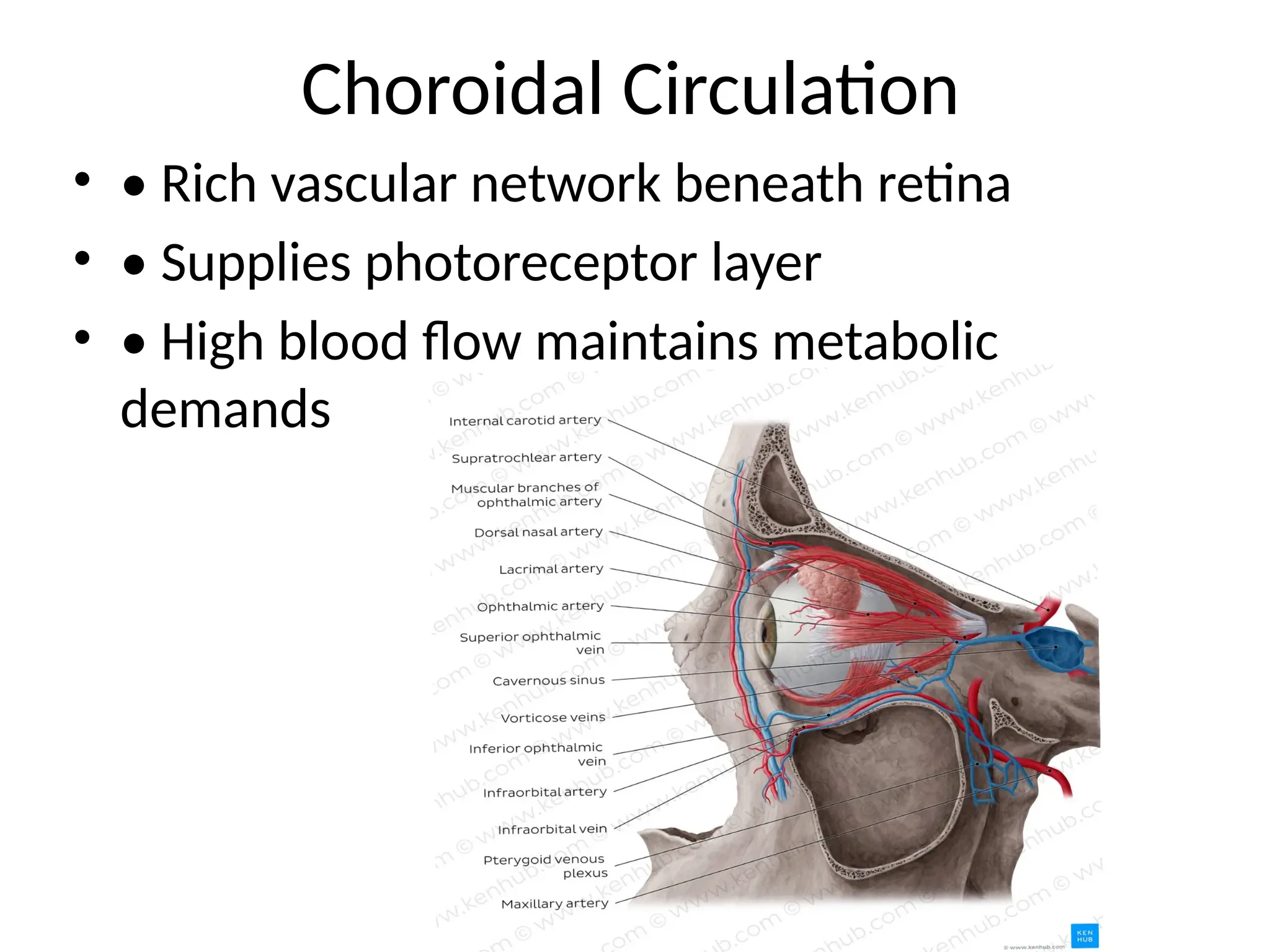
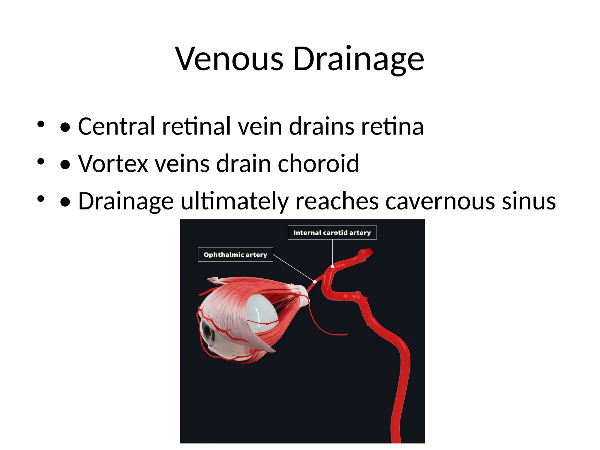
Ophthalmic Artery
• •Main arterial source to the eyeball
• • Branch of the internal carotid artery
• • Enters orbit through optic canal
• • Gives several branches supplying ocular
structures
4.
Central Retinal Artery
•• Branch of ophthalmic artery
• • Pierces optic nerve ~1 cm behind globe
• • Supplies inner retinal layers
• • End artery—occlusion leads to sudden,
painless vision loss
References
• 1. Gray'sAnatomy: The Anatomical Basis of
Clinical Practice
• 2. Clinical Anatomy by Richard Snell
• 3. Ophthalmology by Yanoff & Duker
• 4. www.aao.org (American Academy of
Ophthalmology)