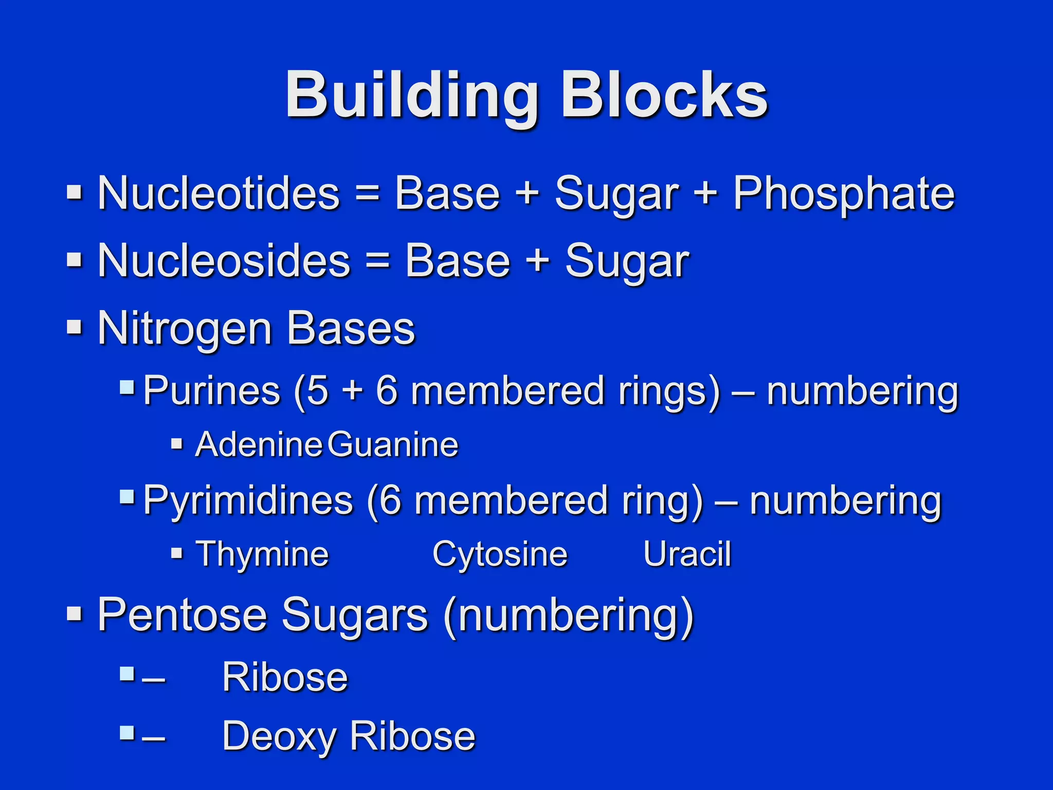
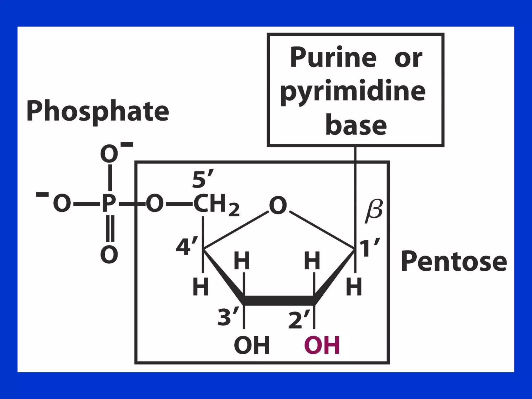
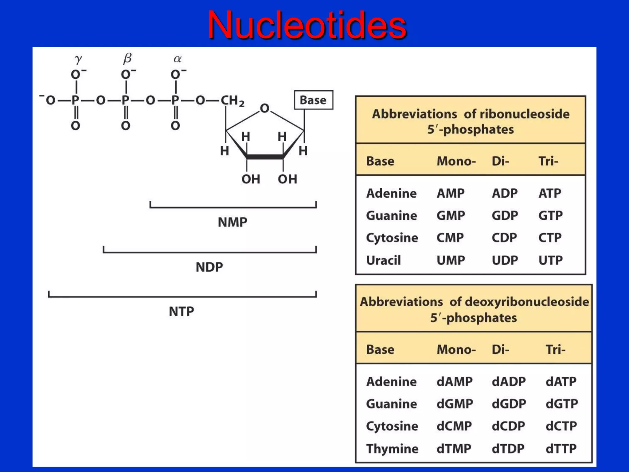
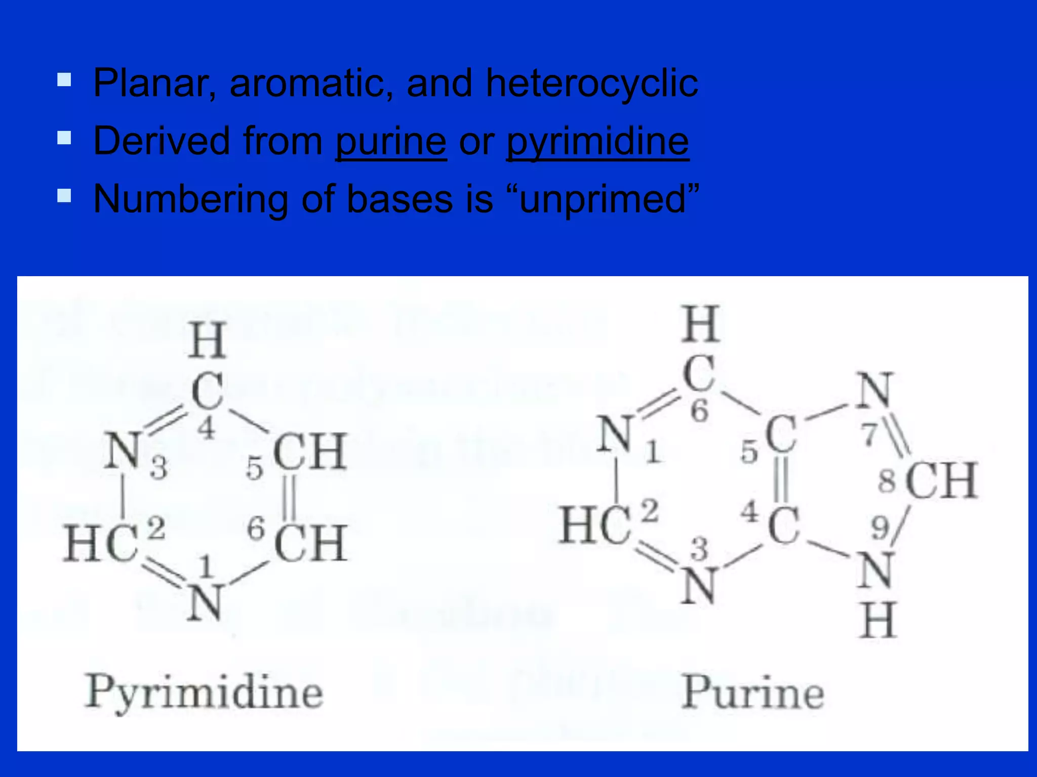
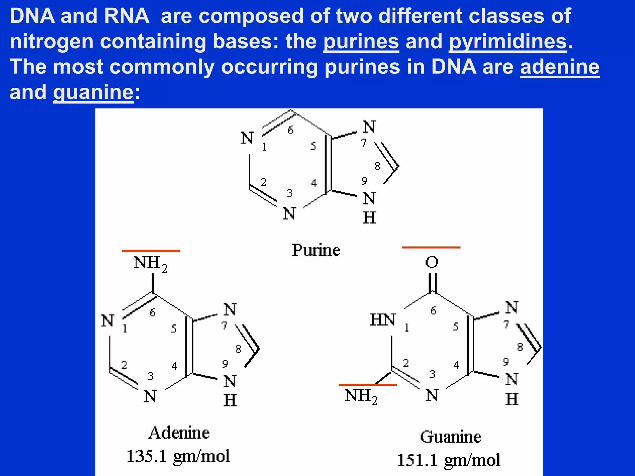
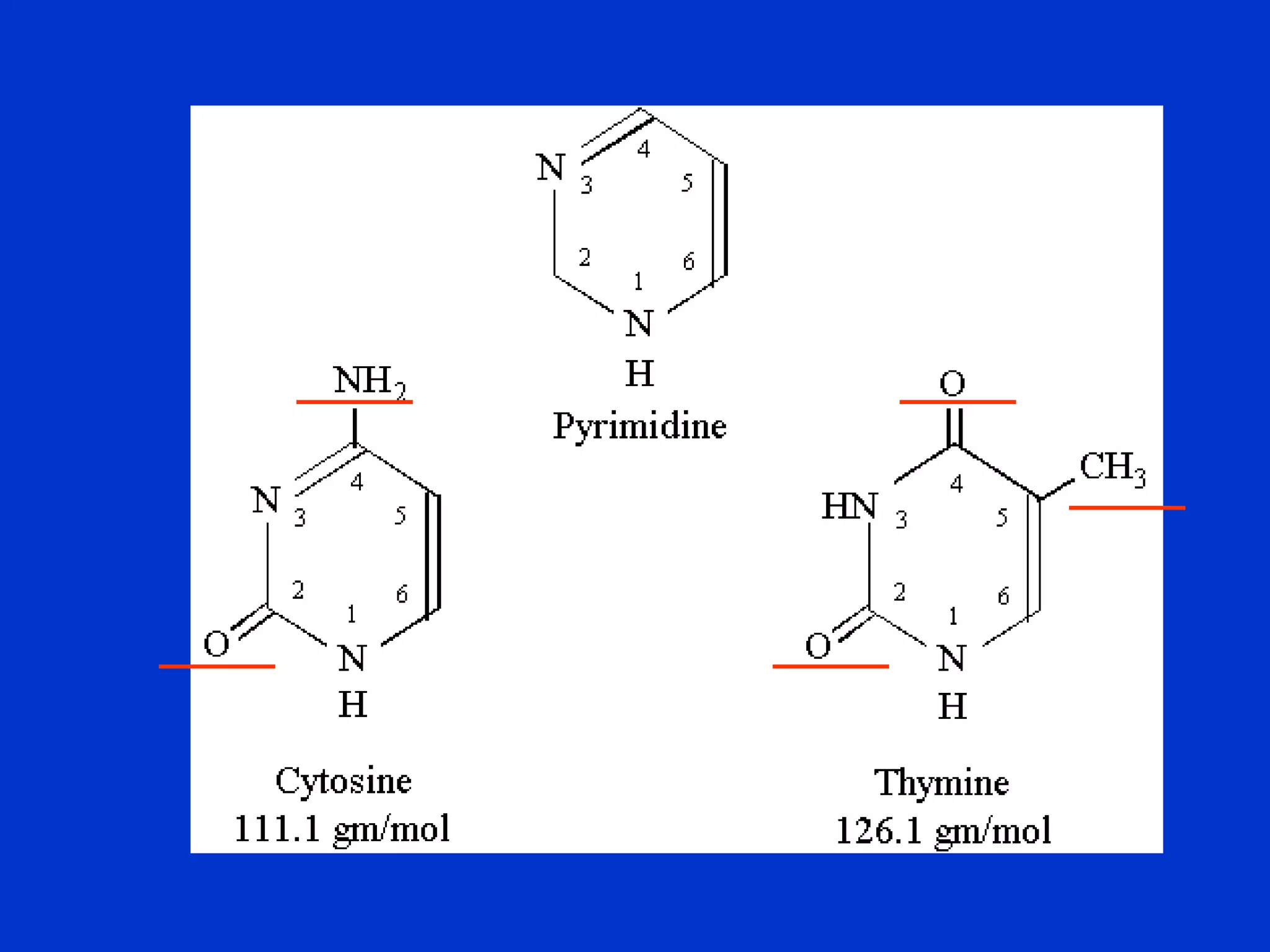
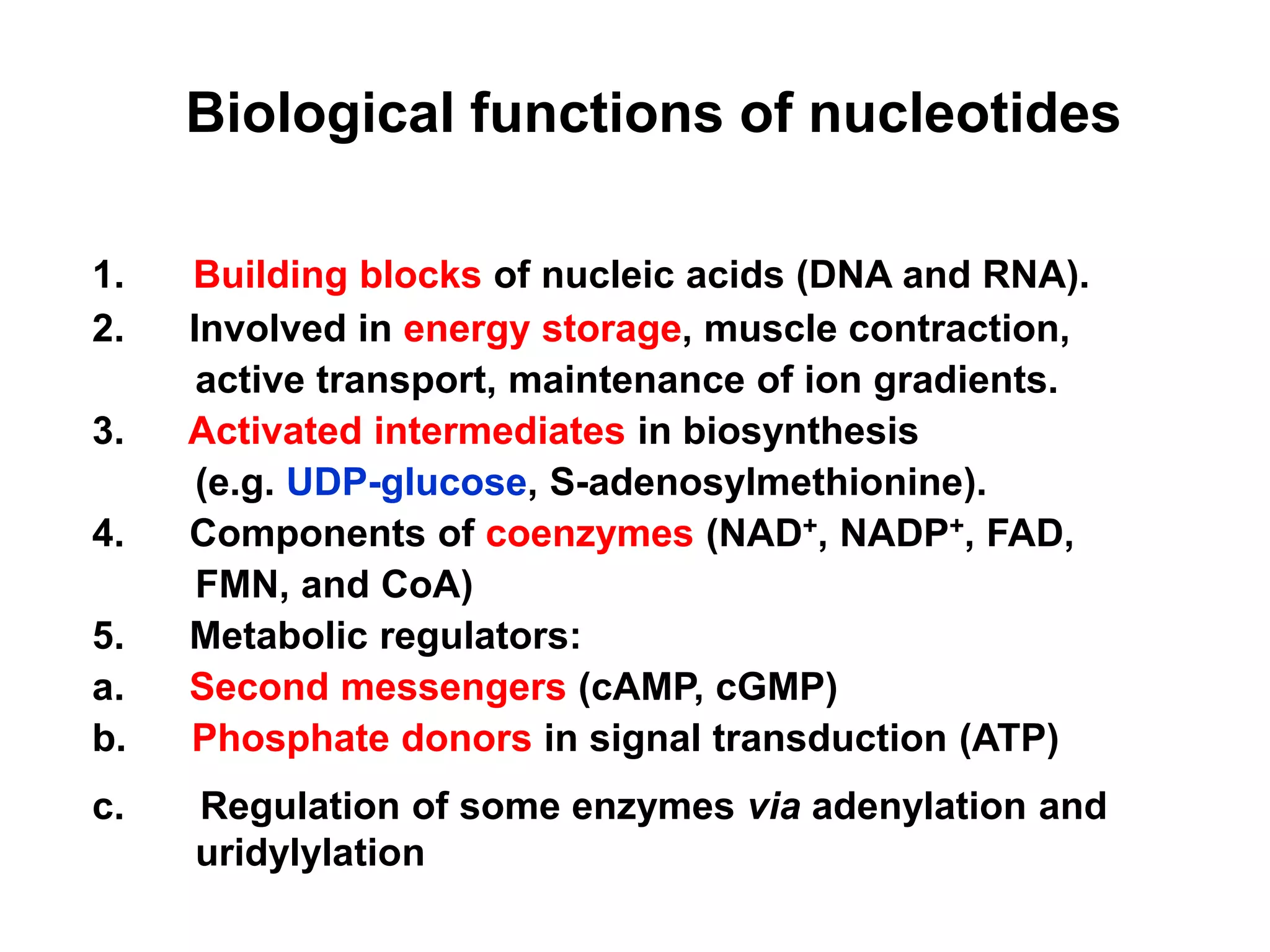
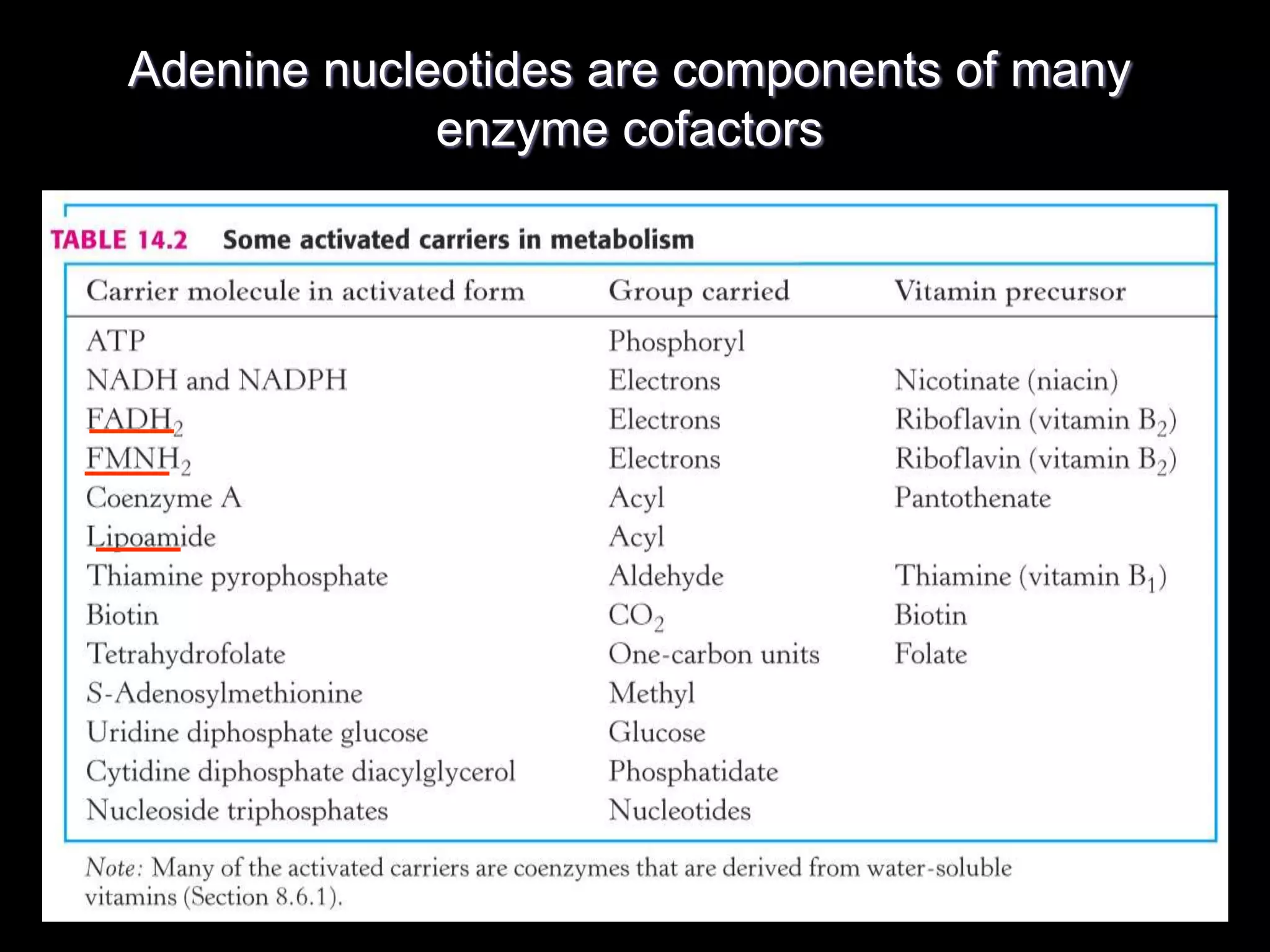
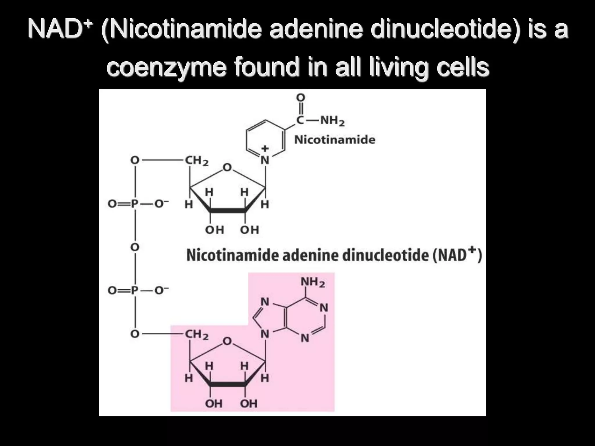
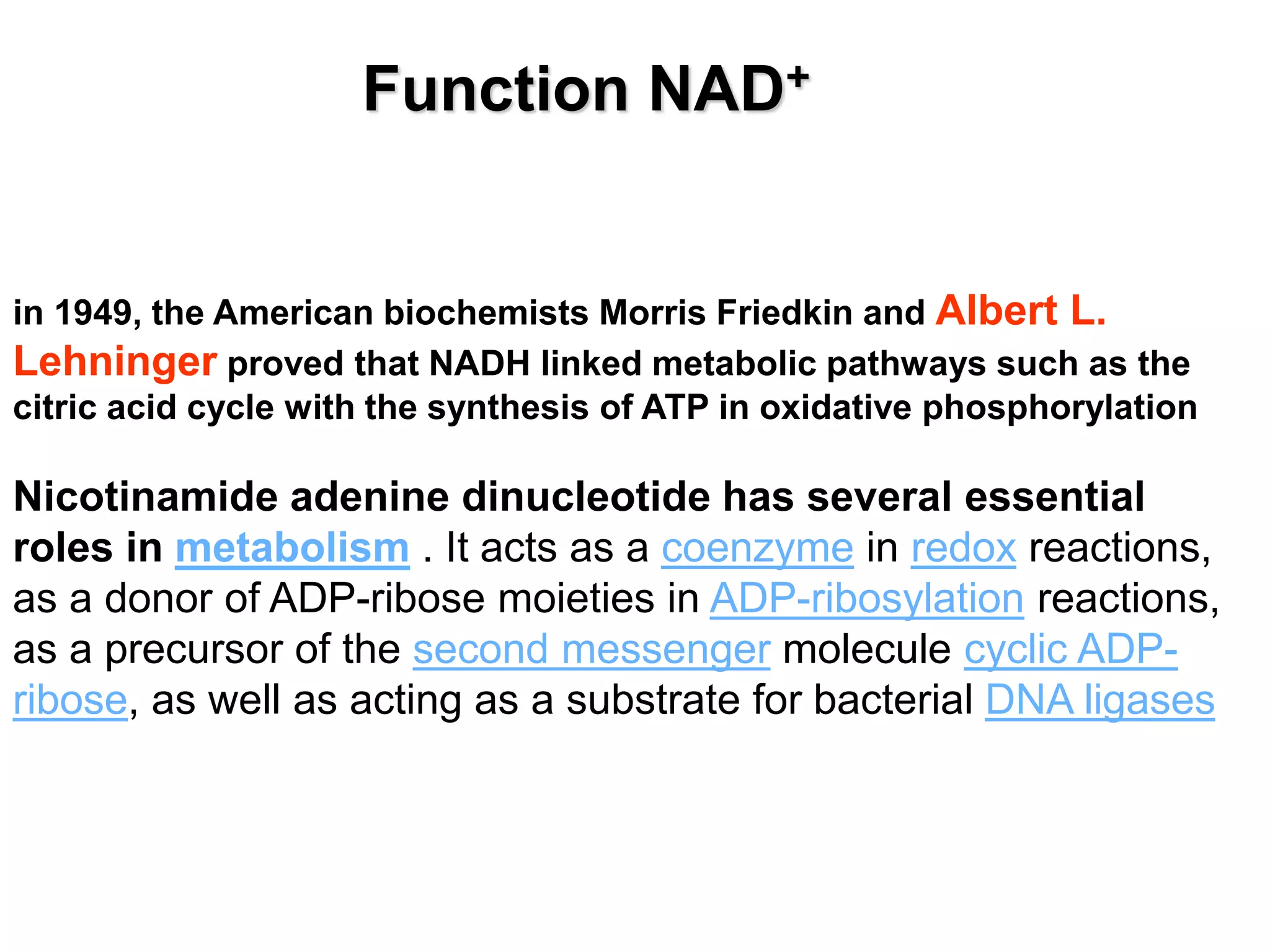
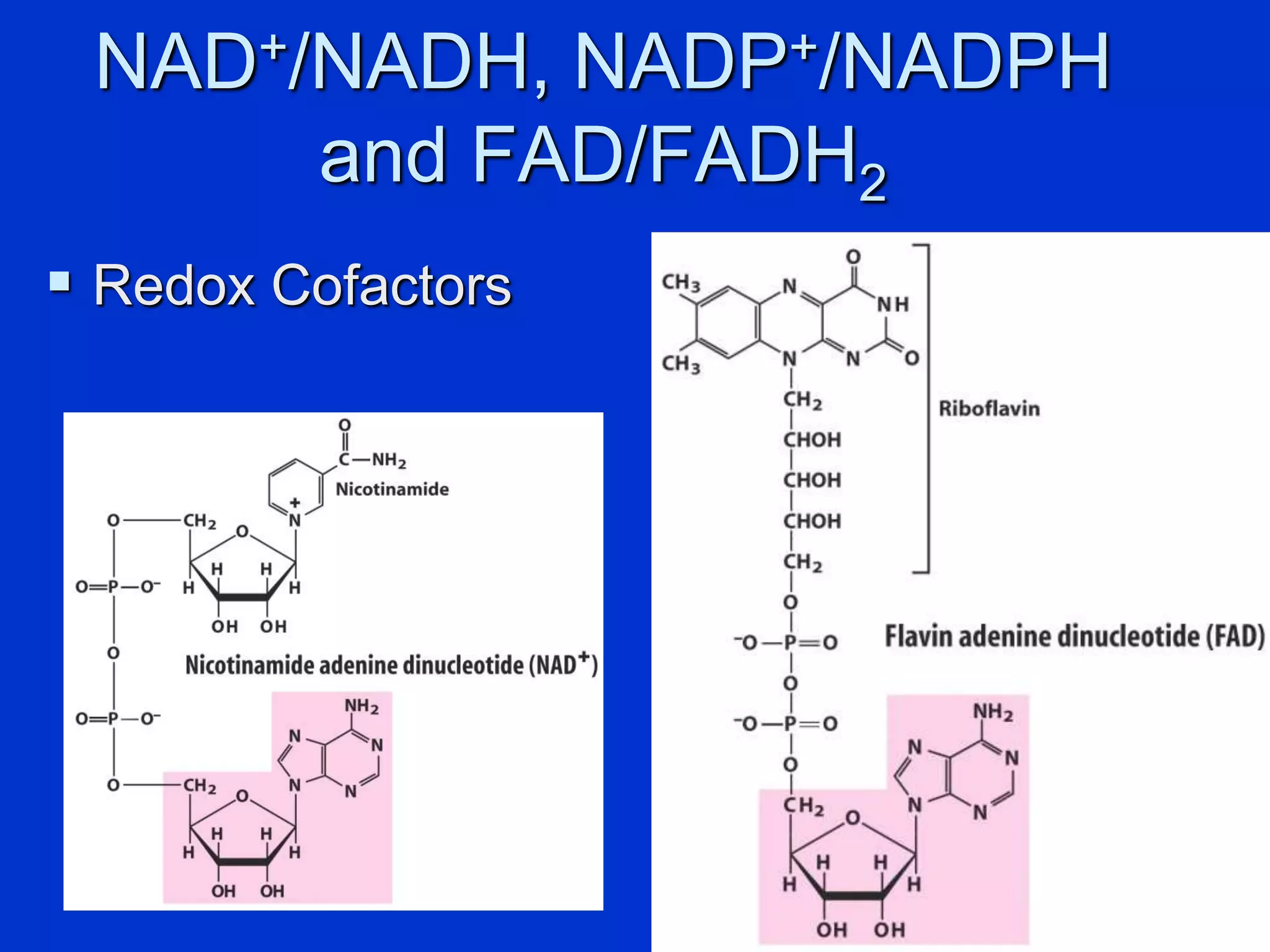
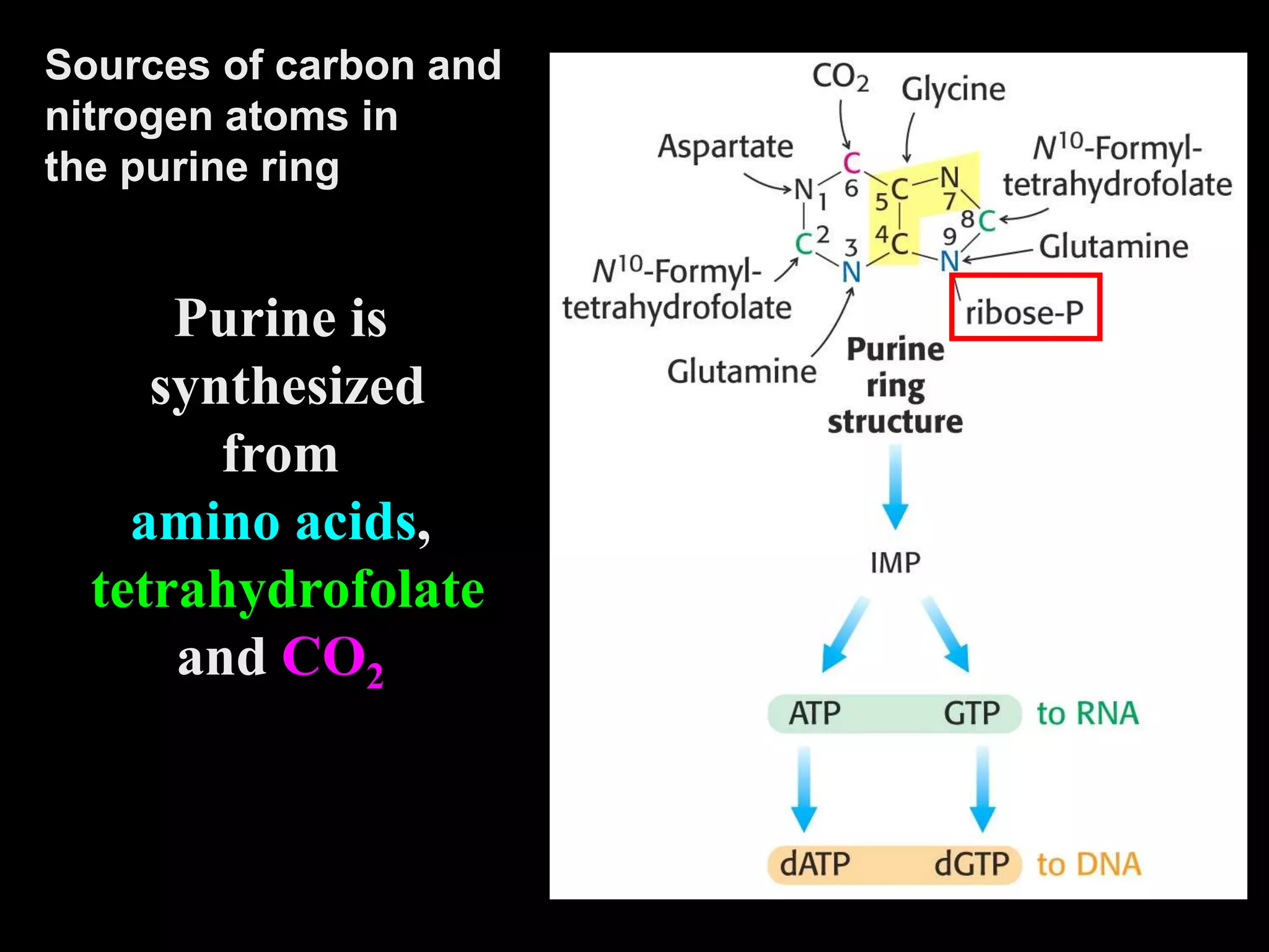
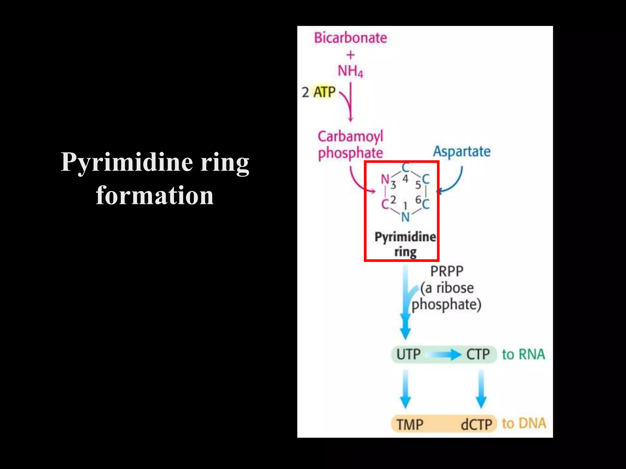
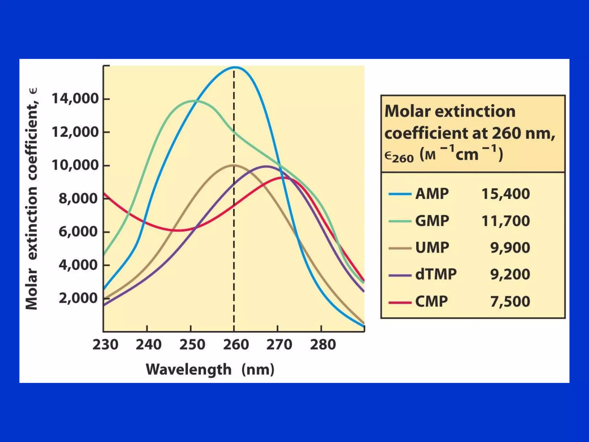
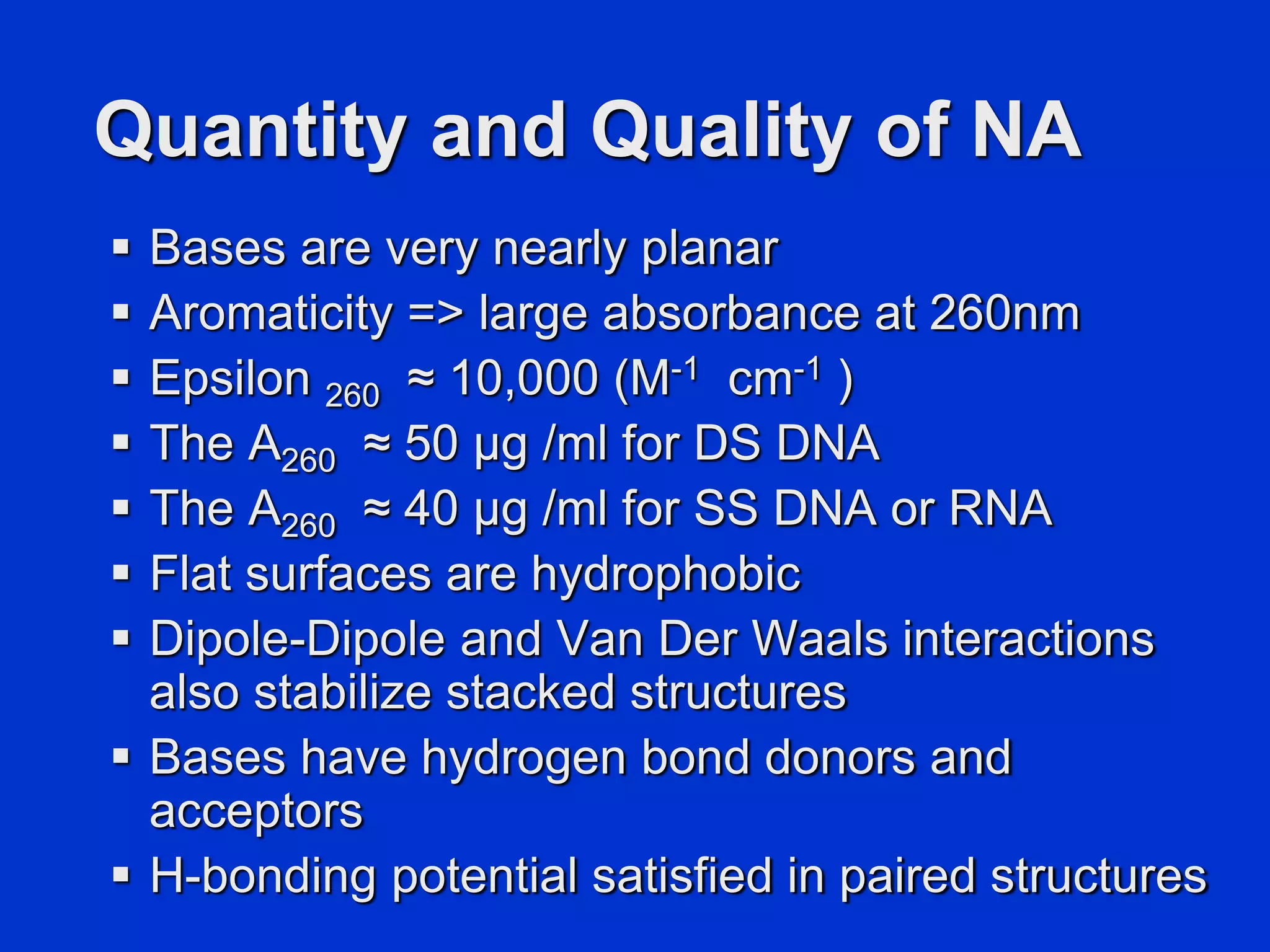
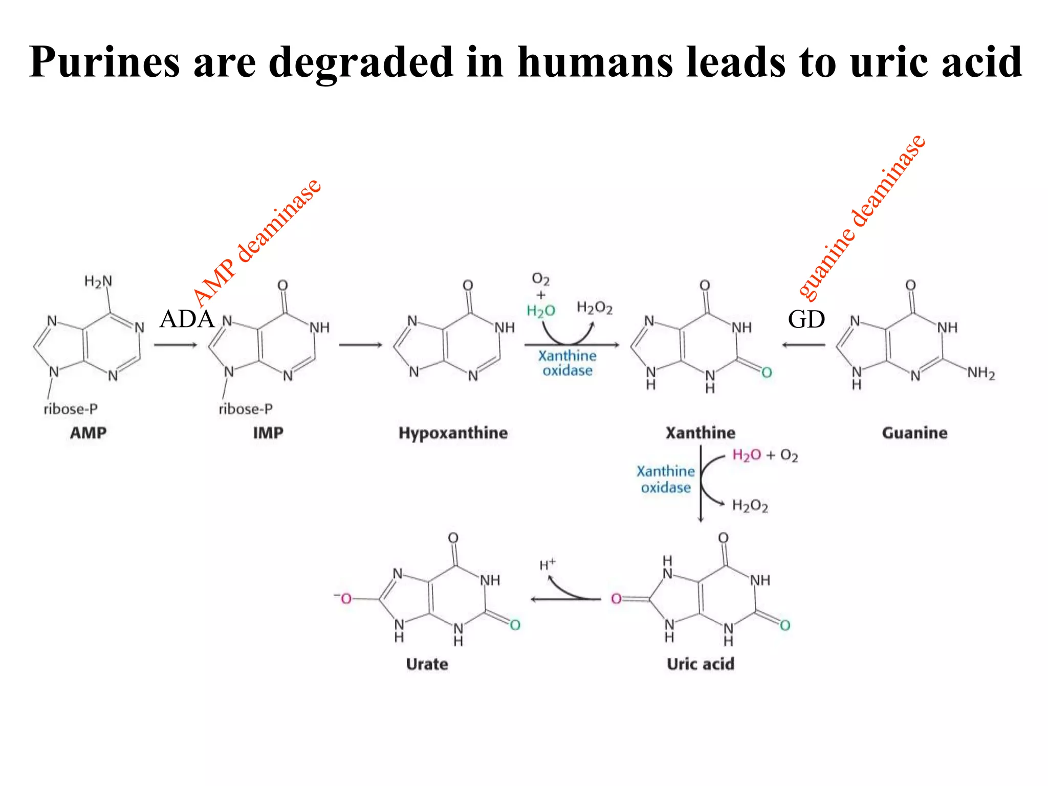
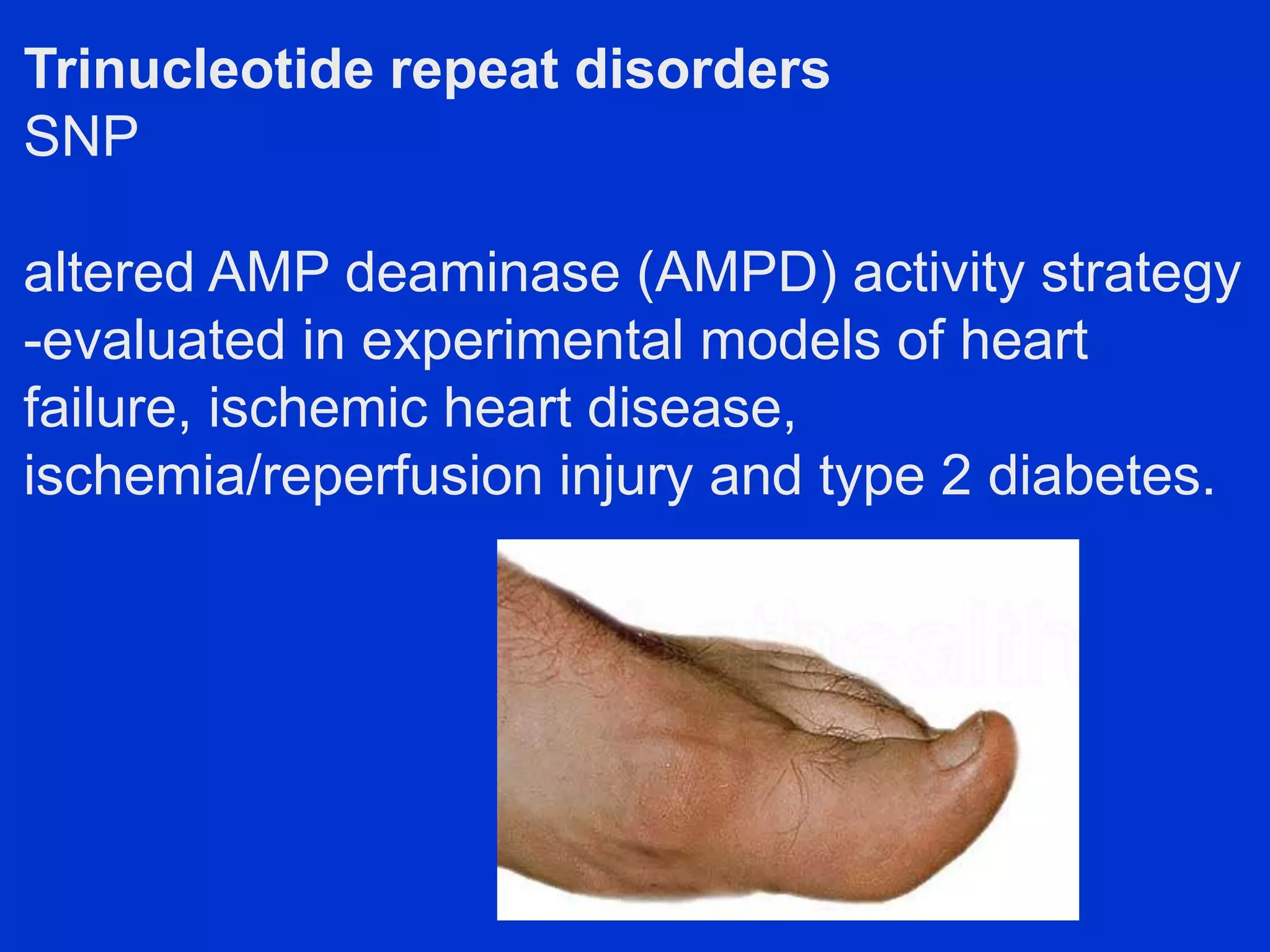
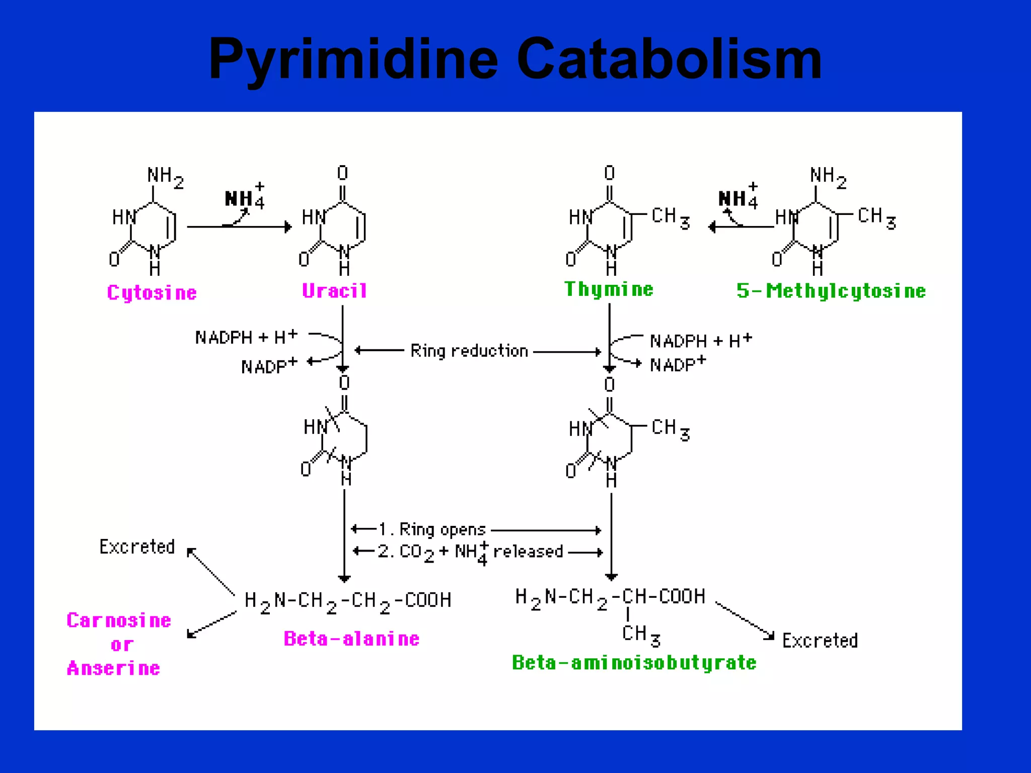
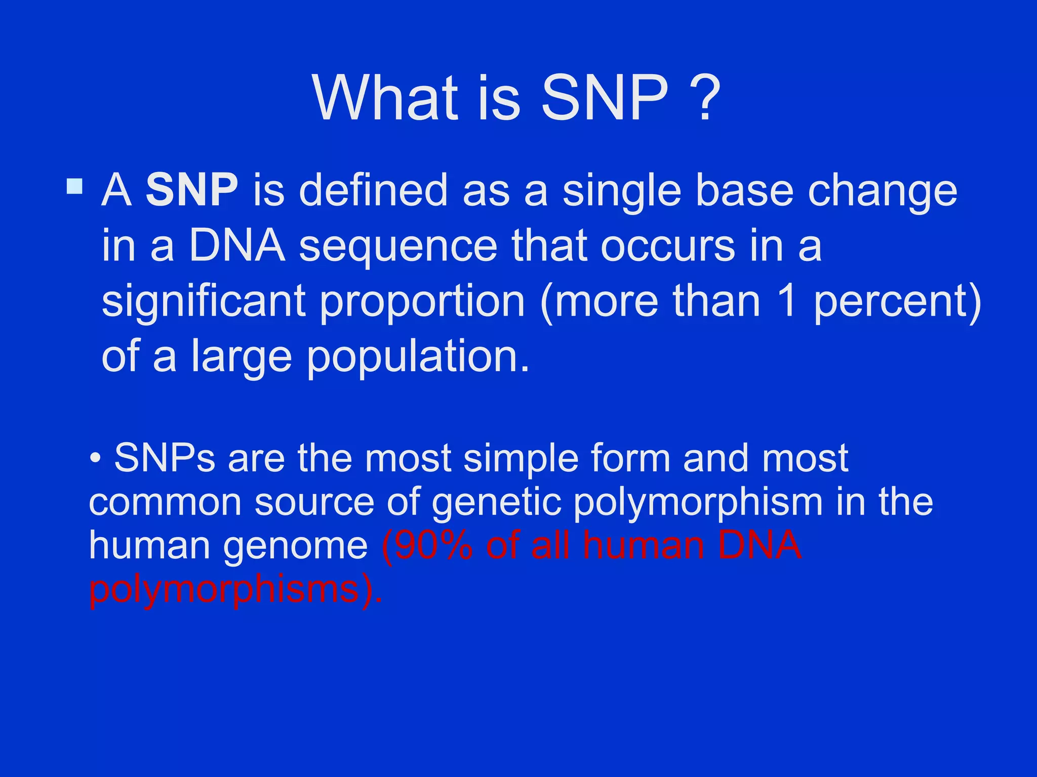
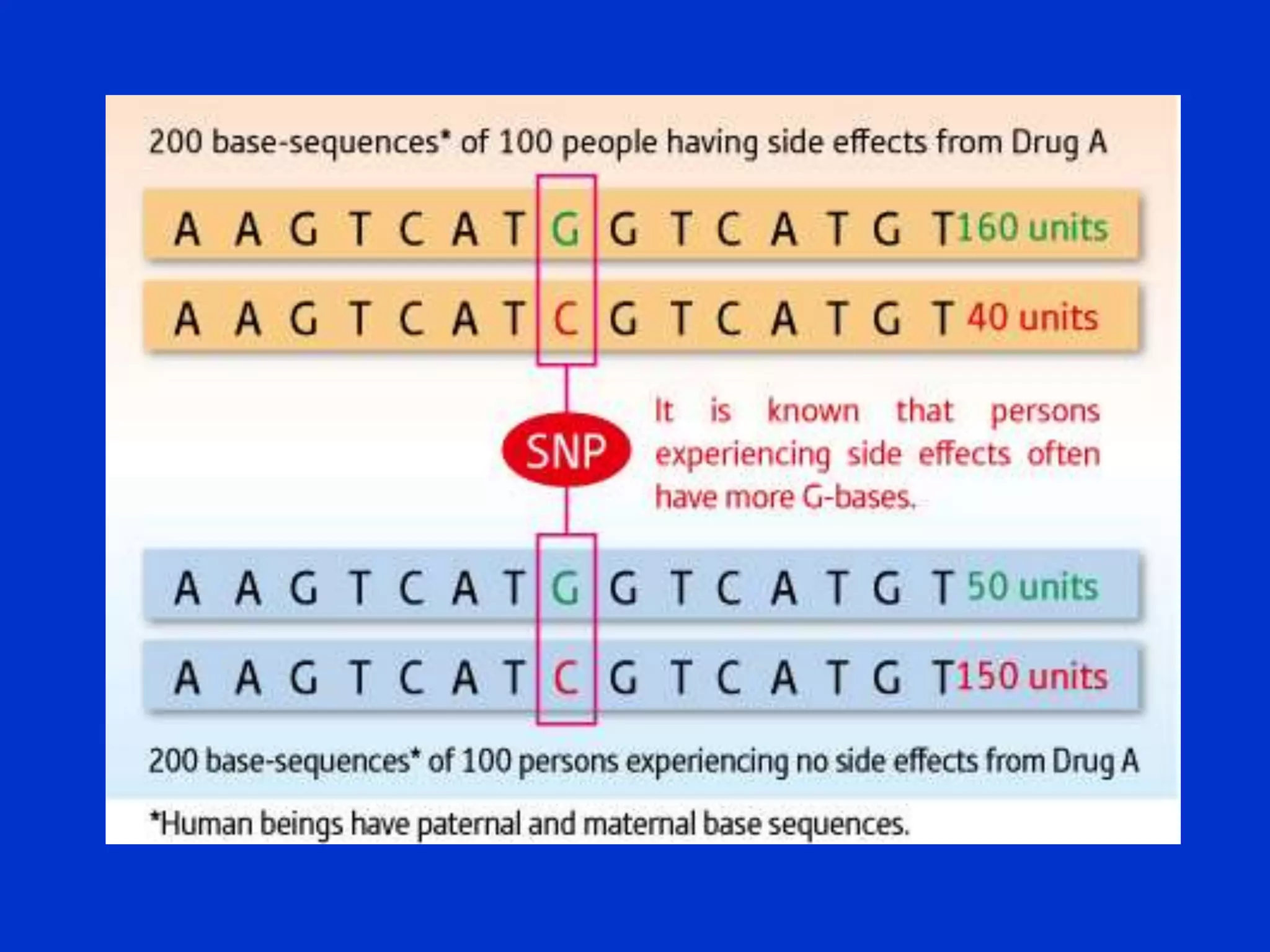
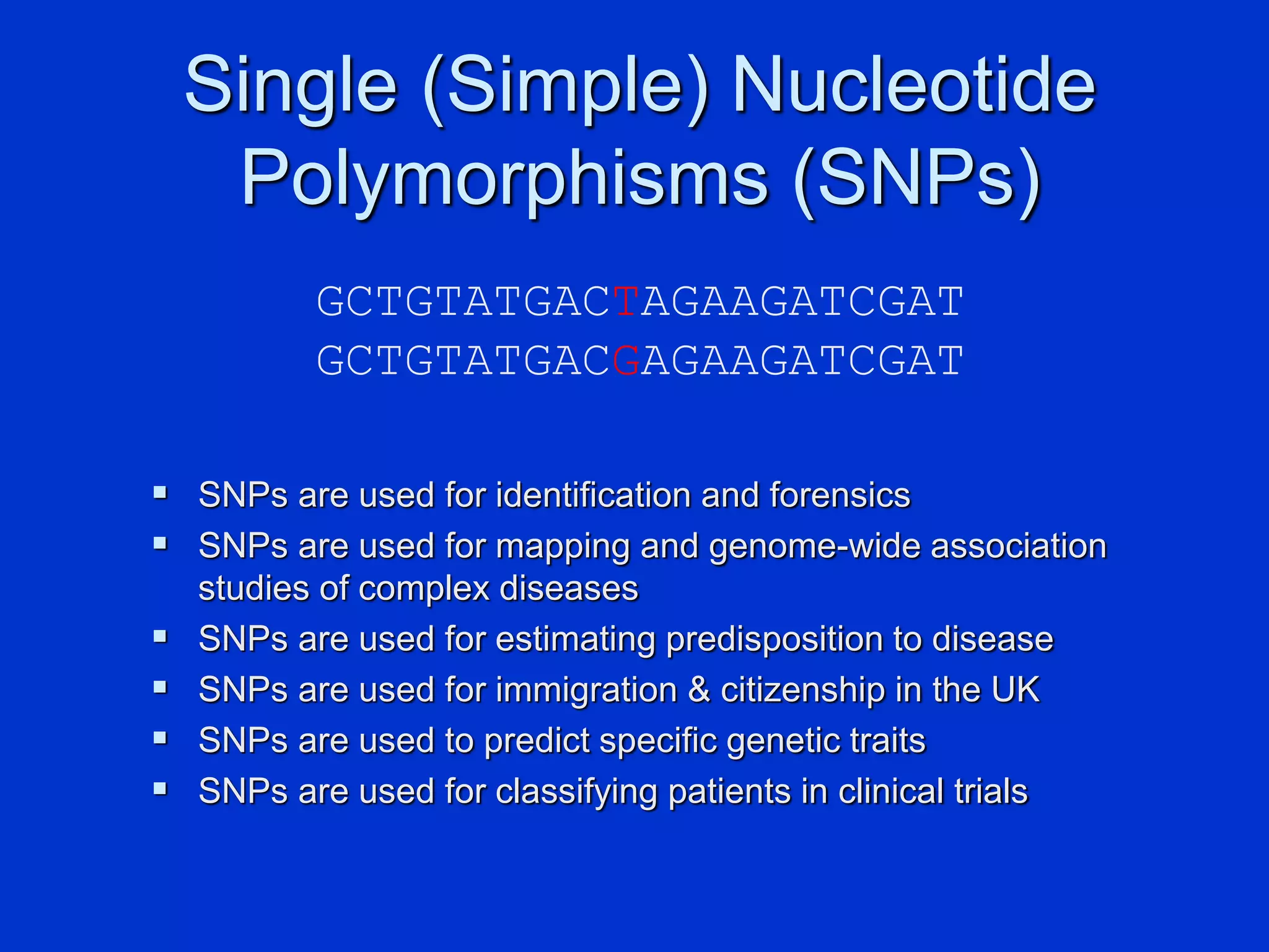
The document provides an overview of the structure and function of nucleic acids, specifically purines, pyrimidines, nucleosides, and nucleotides, detailing their roles in molecular biology and genetics. It elaborates on the biological functions of nucleotides, including energy transfer, biosynthesis, and metabolic regulation, as well as discussing specific nucleotides like ATP, GTP, UTP, and CTP. Additionally, it addresses the metabolism of purines and pyrimidines, significance of single nucleotide polymorphisms (SNPs), and their applications in genetics and medicine.

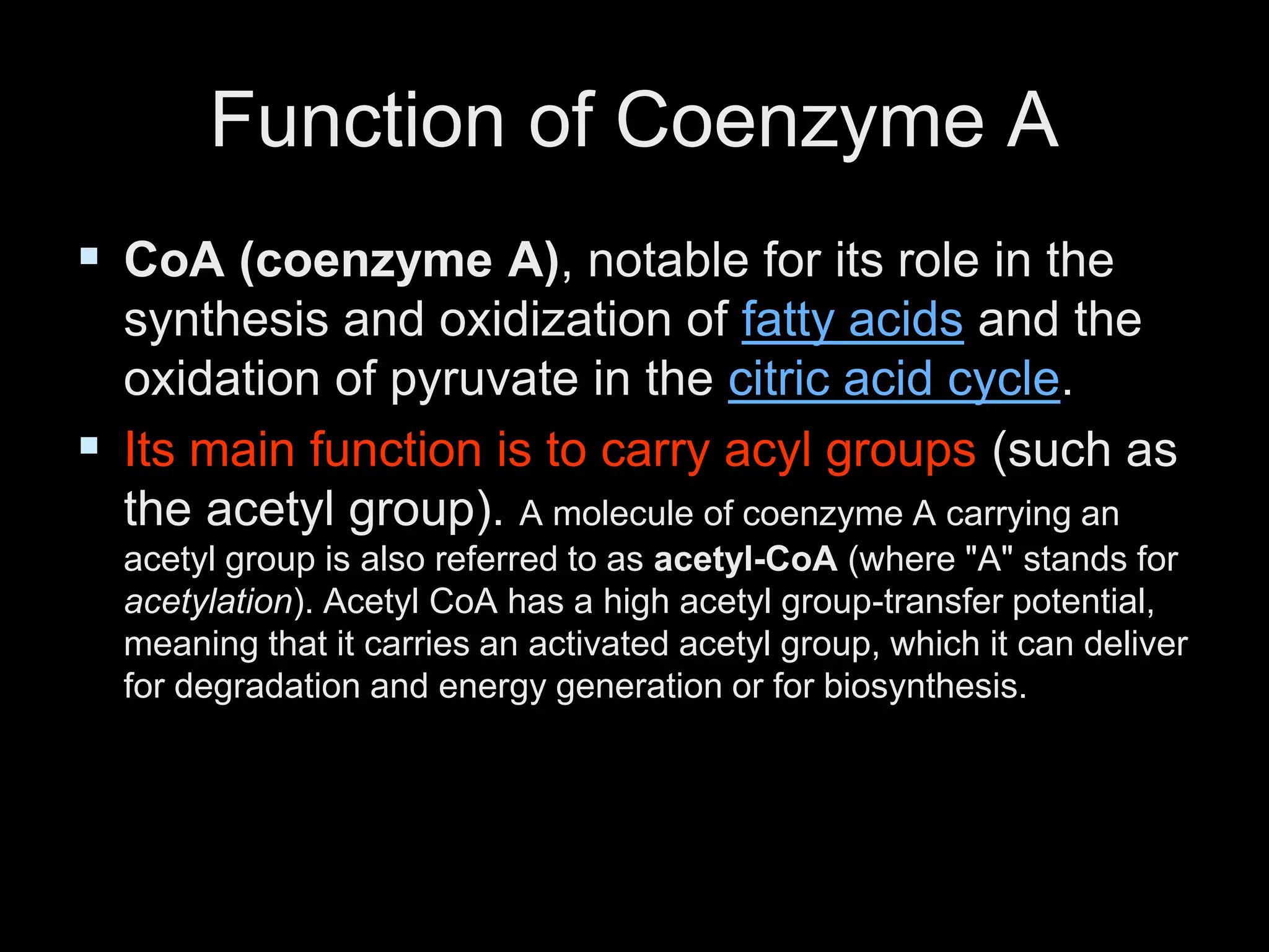


![SHS_Core_CAE_Q3_LE1 FOR THIRD [FINAL].pdf](https://cdn.slidesharecdn.com/ss_thumbnails/shscorecaeq3le1final-251116055110-e3081055-thumbnail.jpg?width=640&height=640&fit=bounds)


