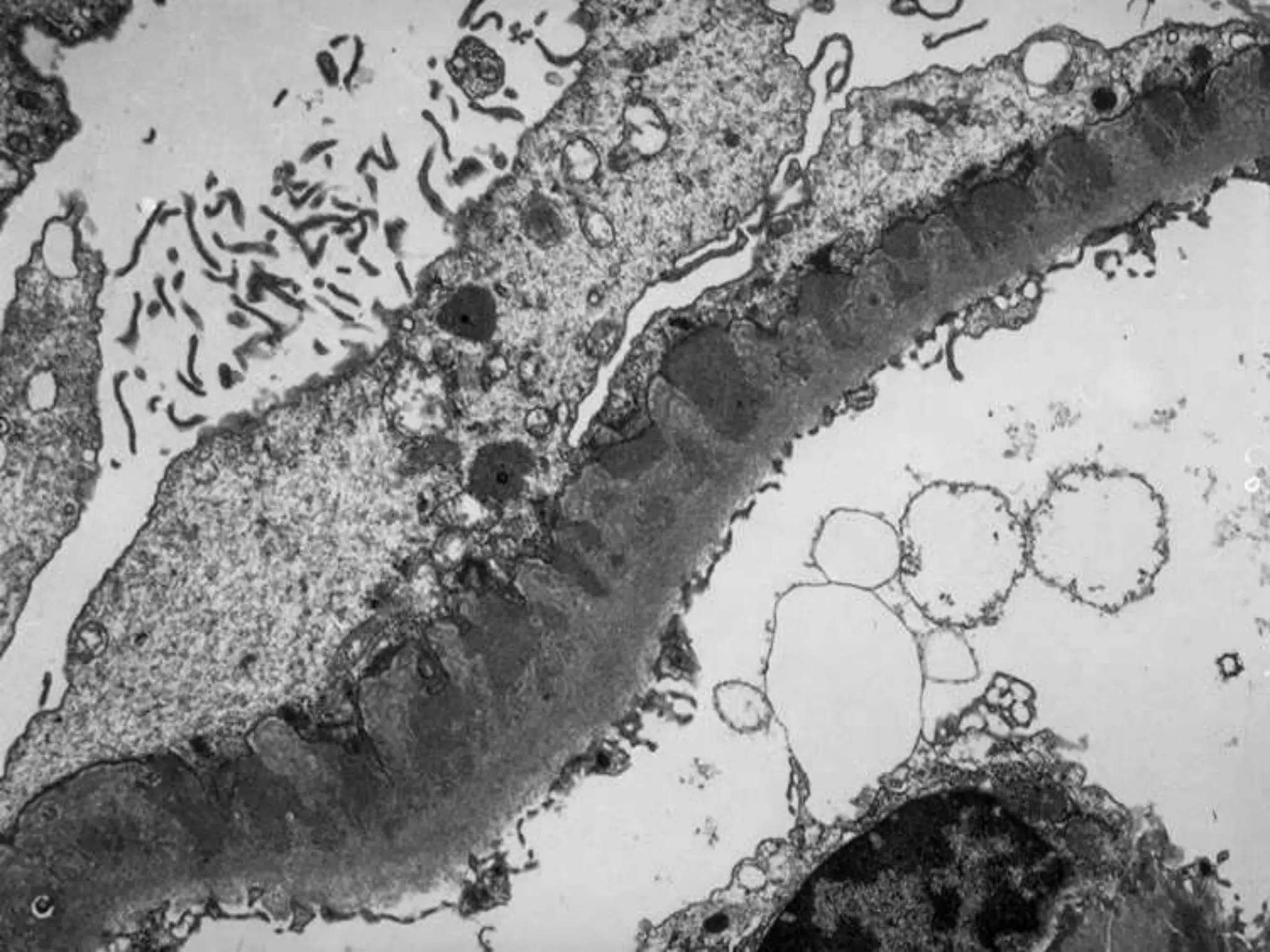
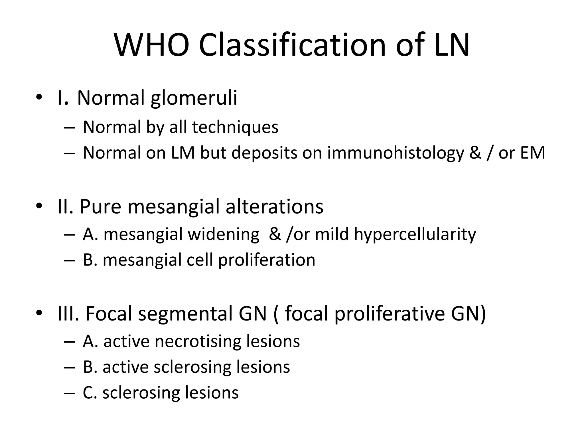
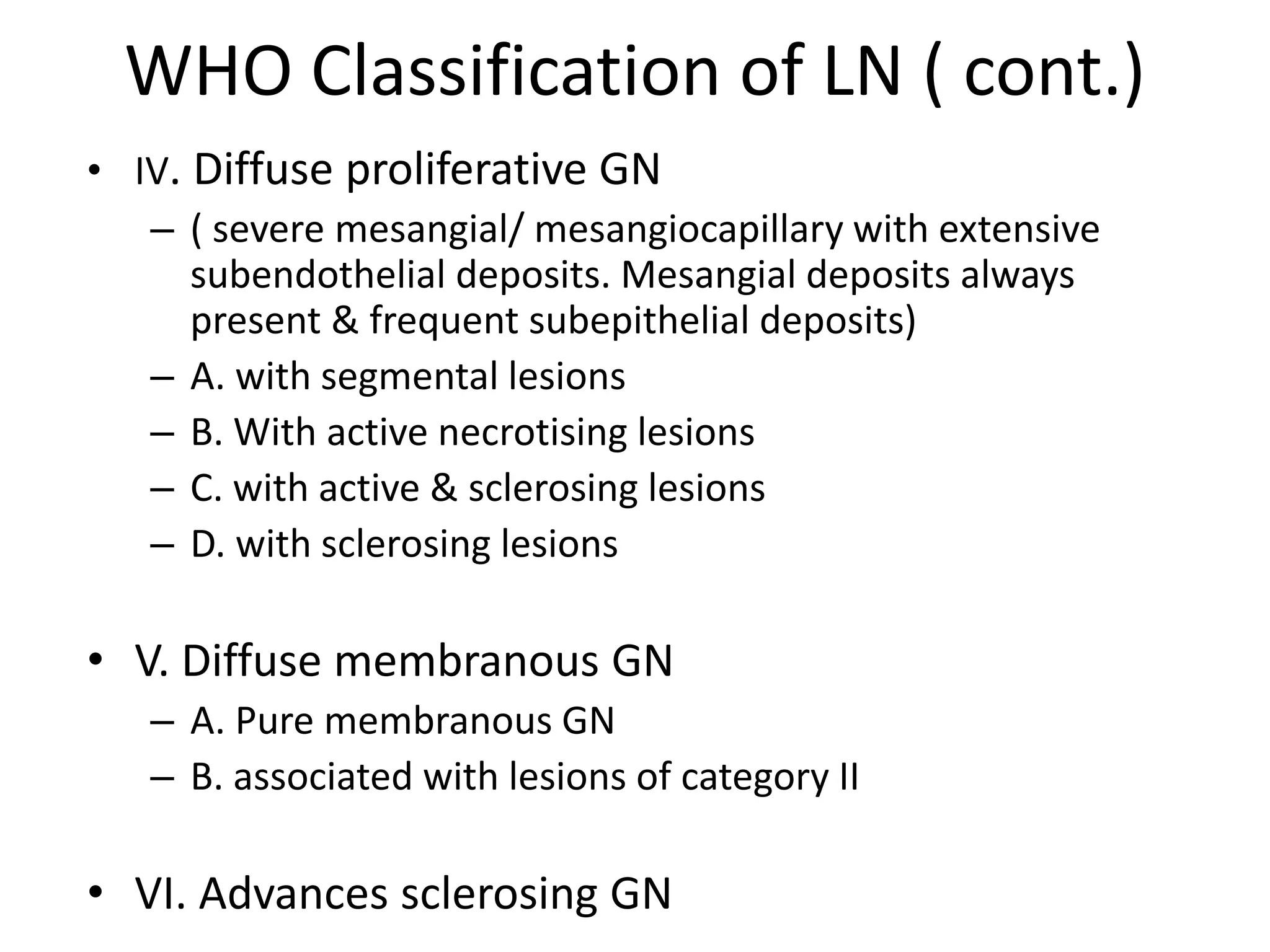
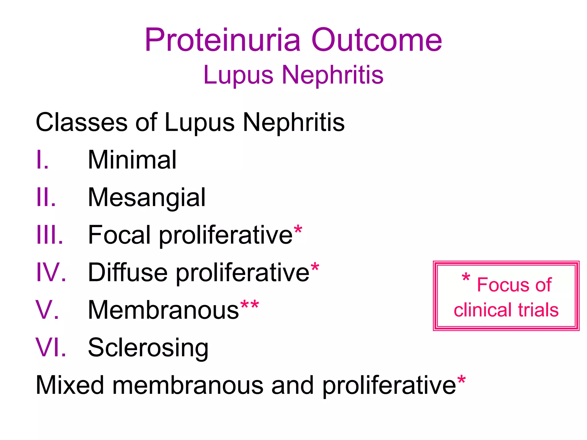
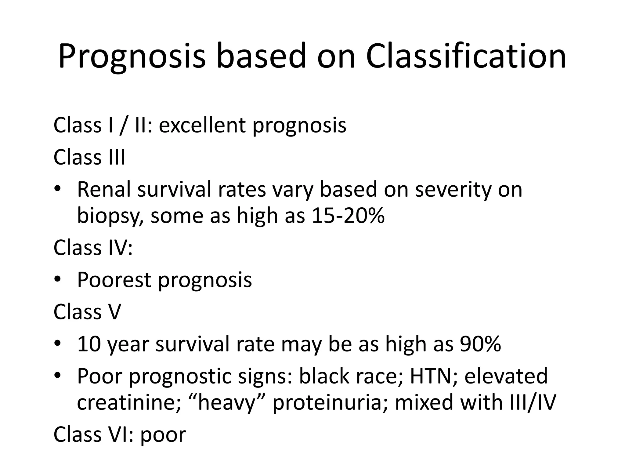
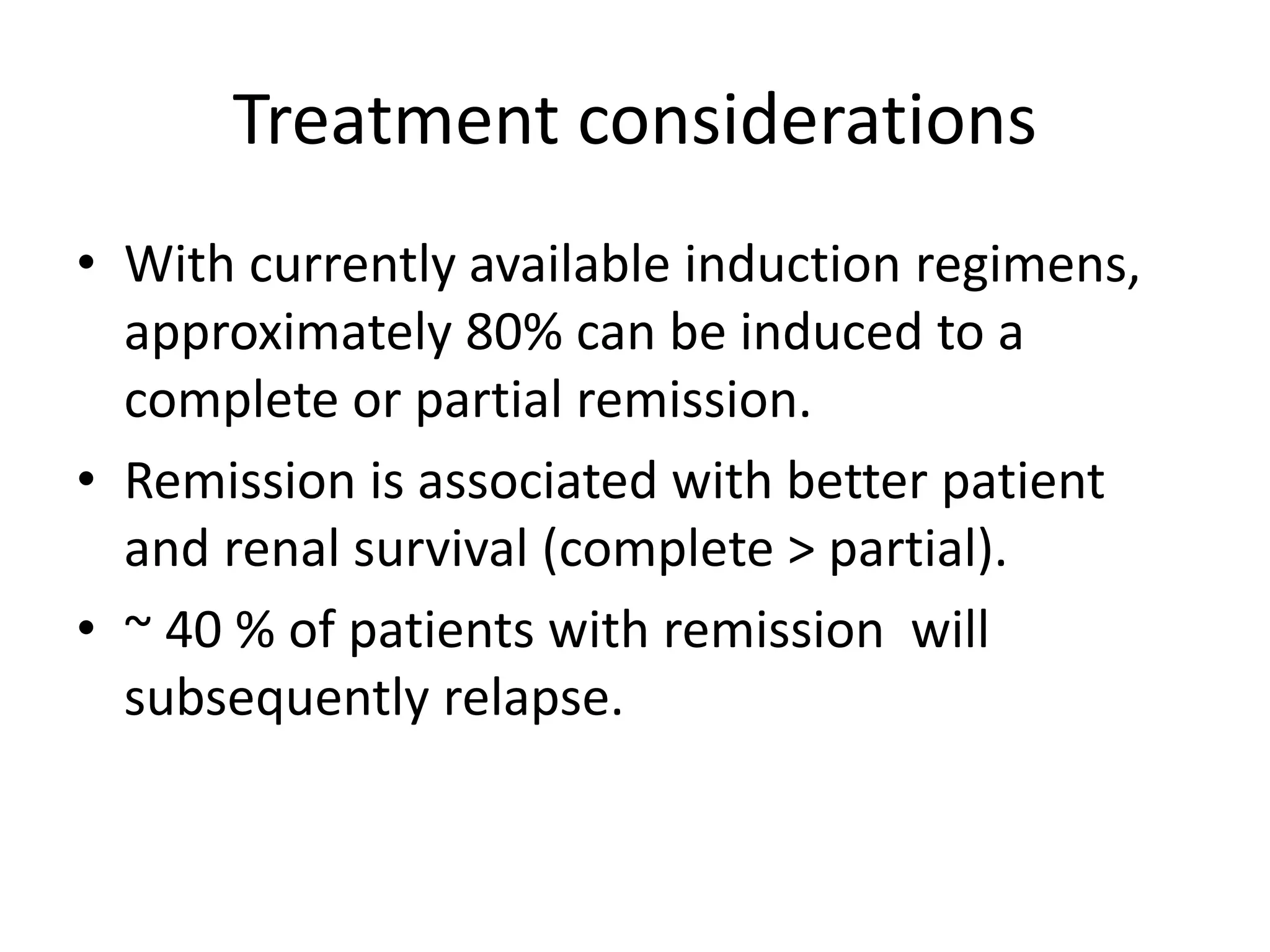
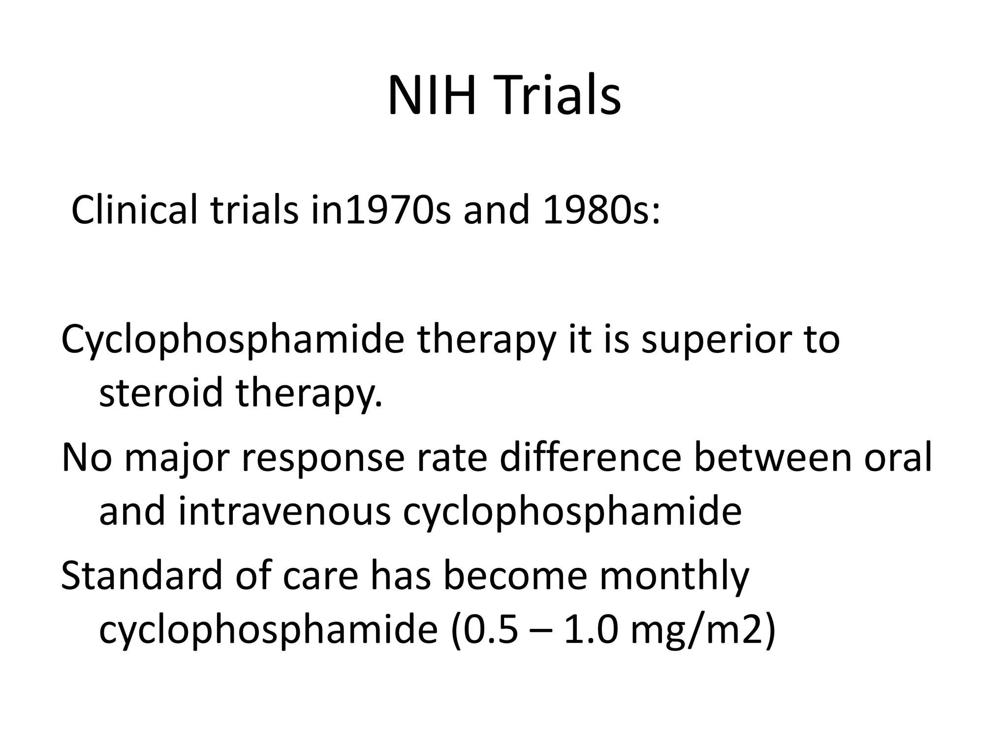
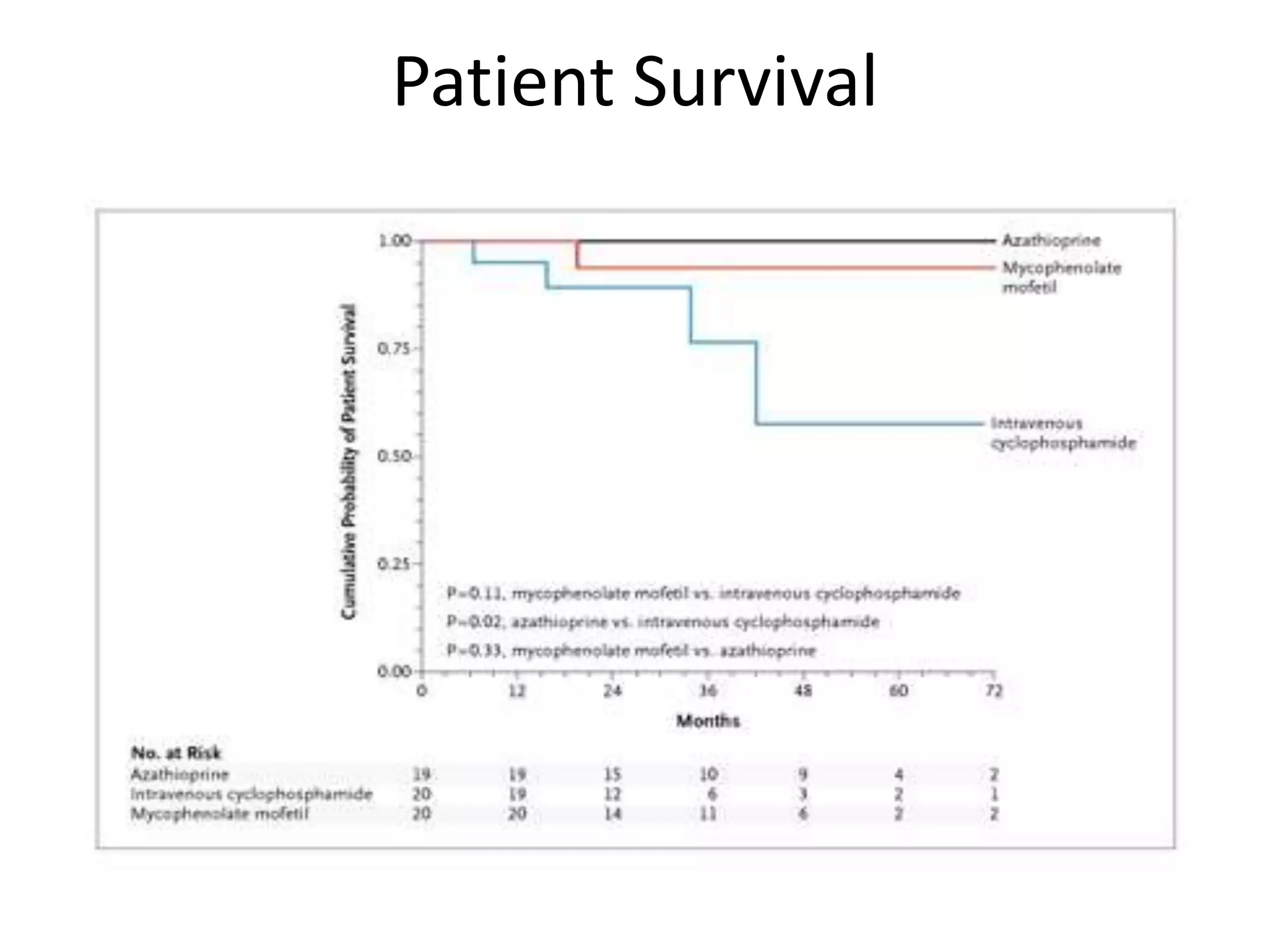
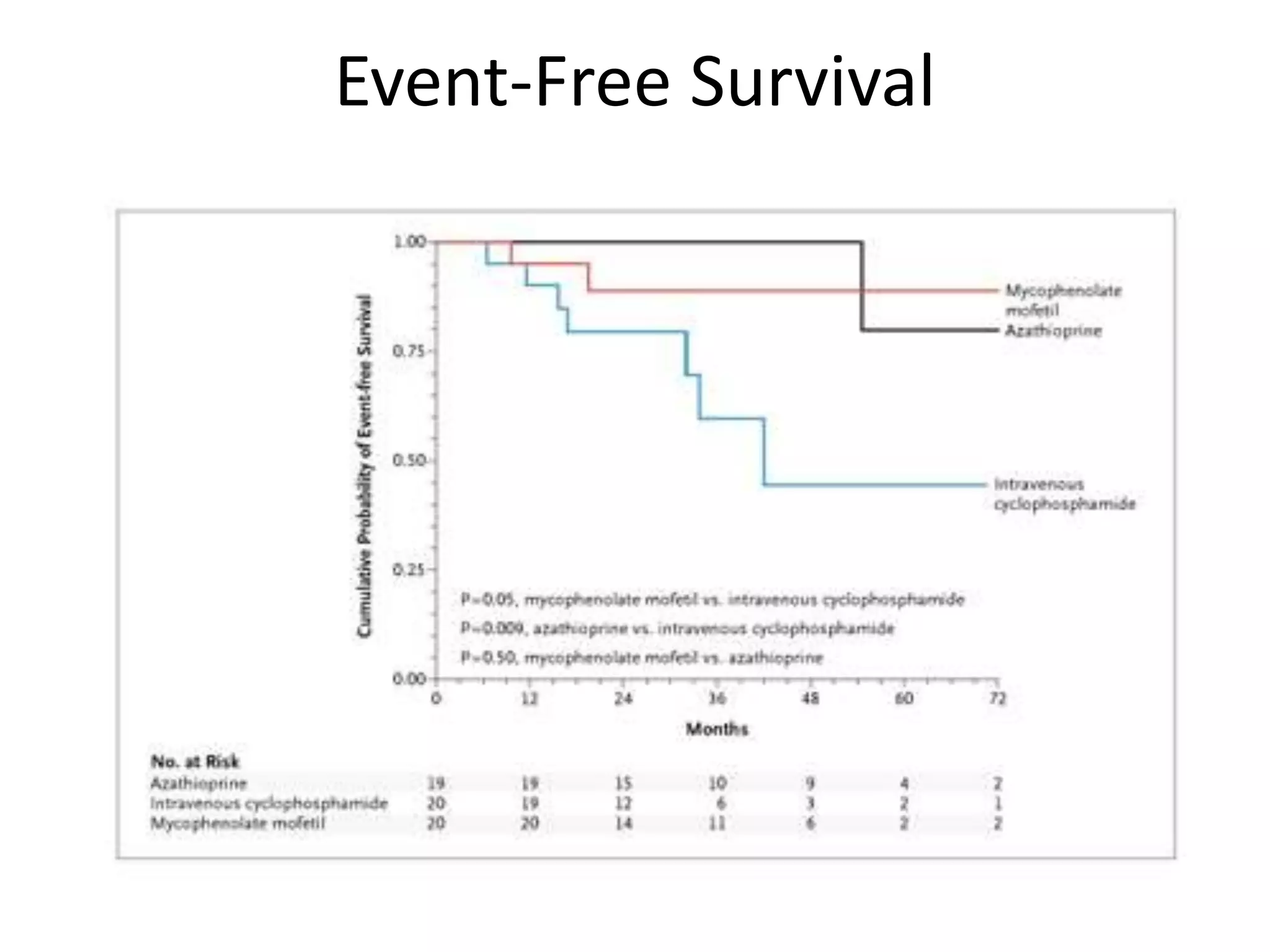
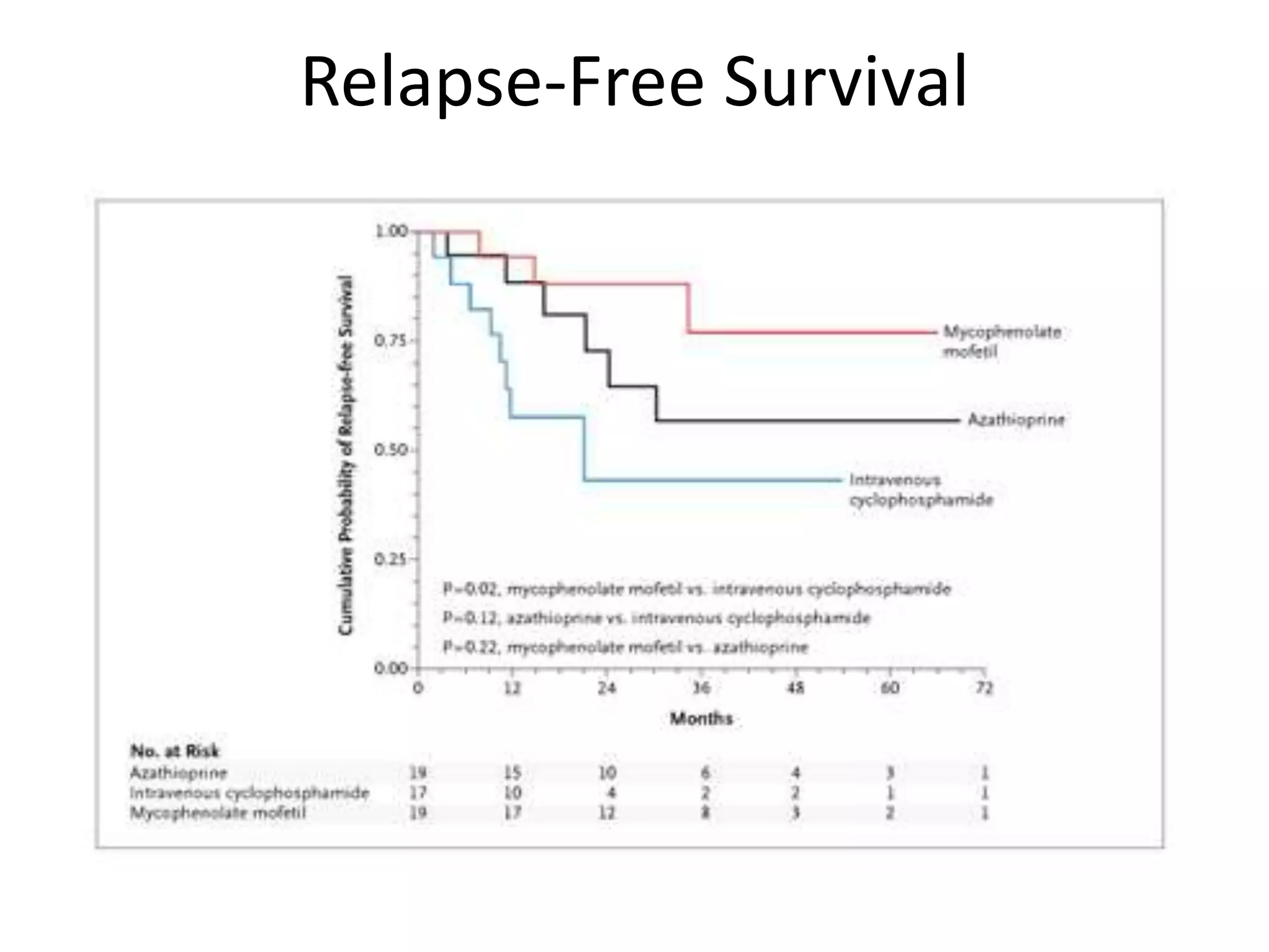
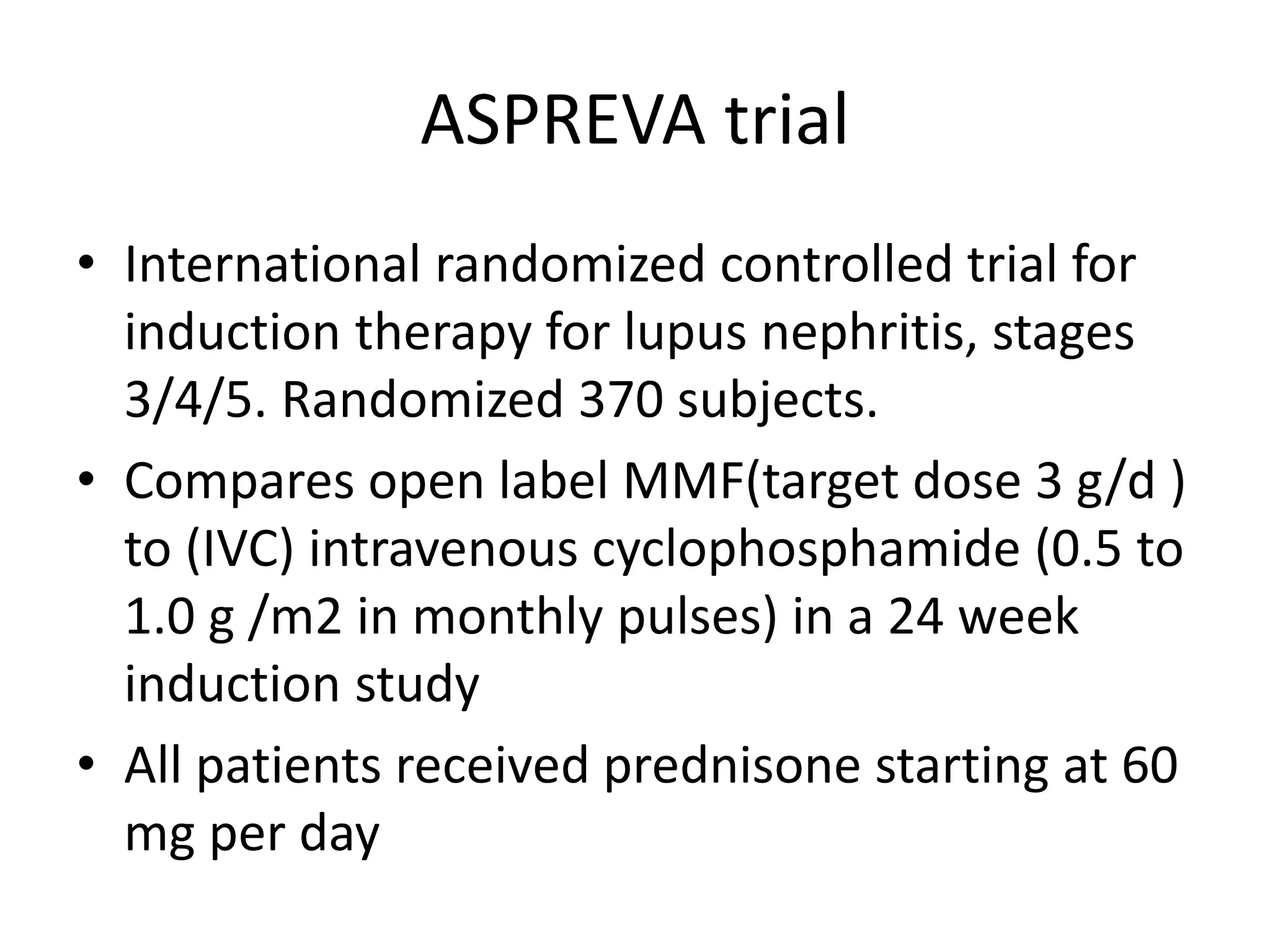
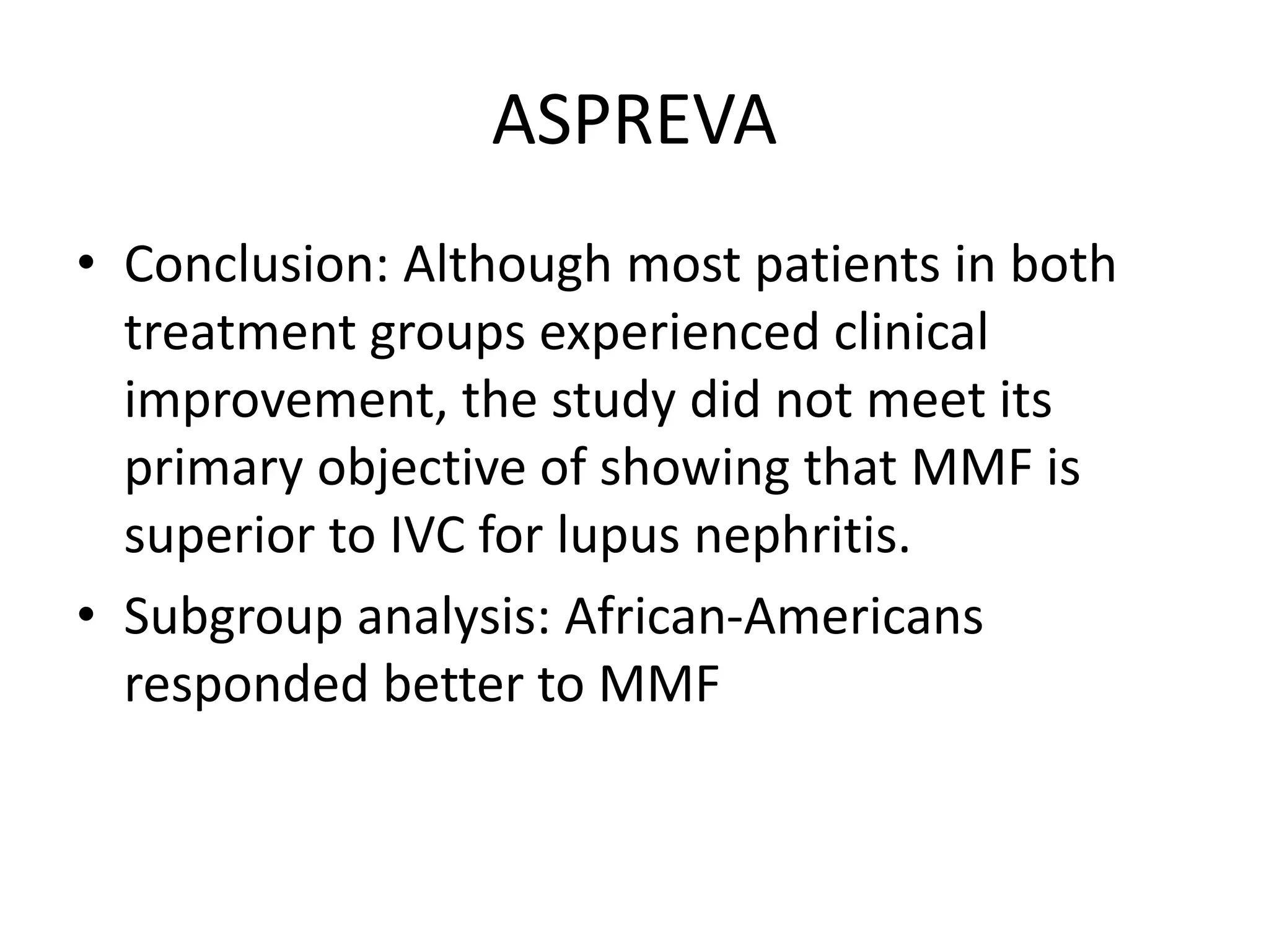
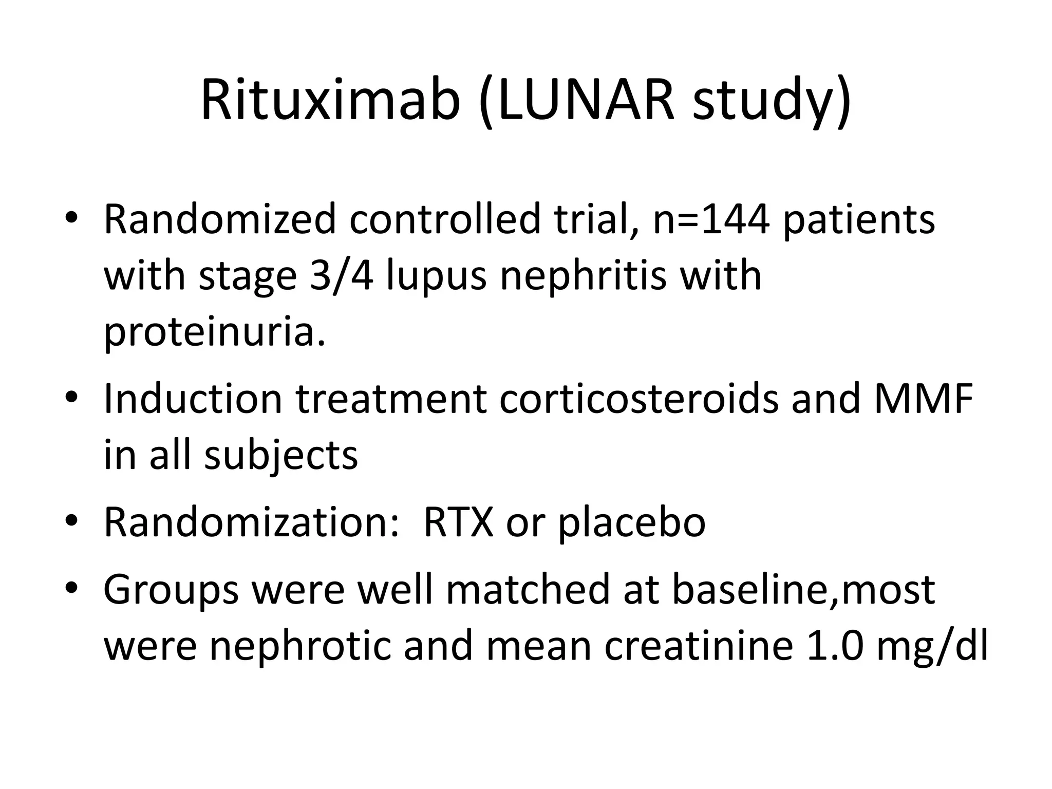
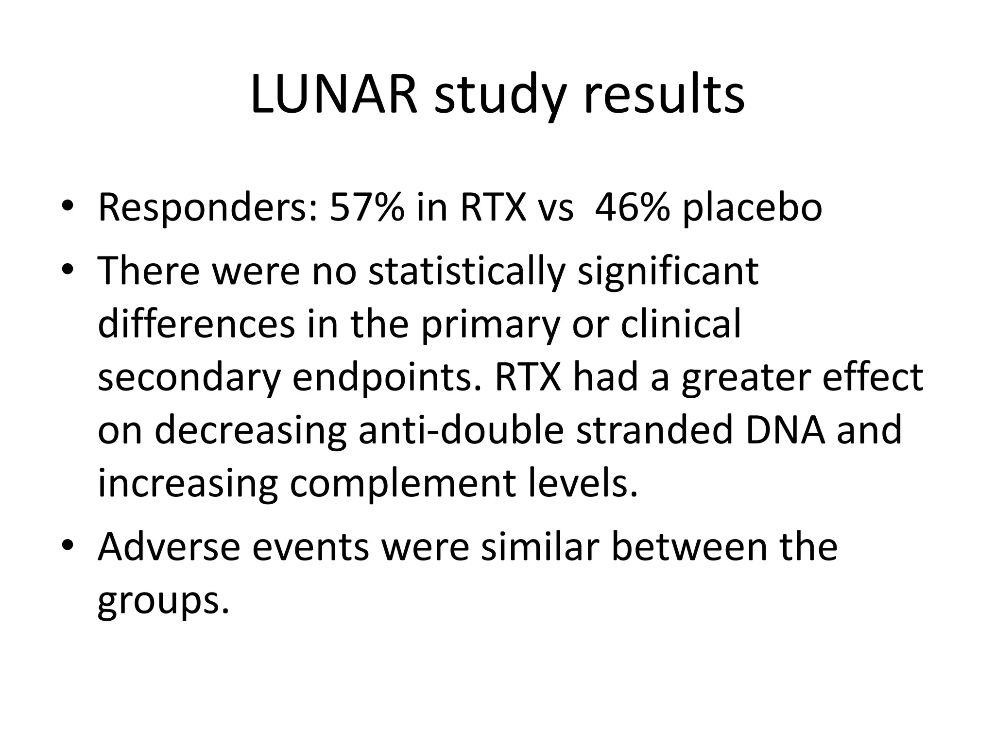
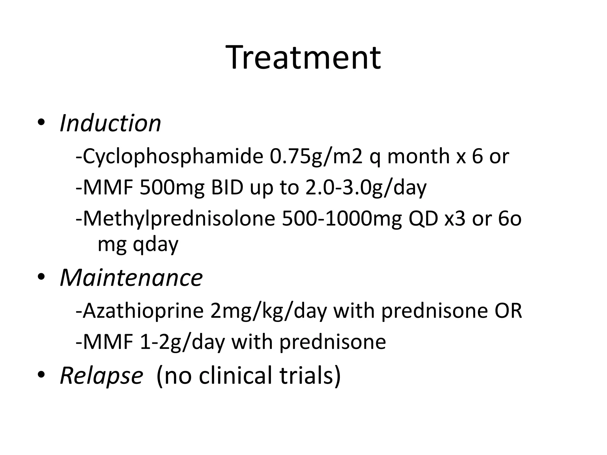
Lupus Nephritis Update 2010
Review of recent treatment trials
Patient example
• 34 year-old unemployed, uninsured, poverty
level income, African-American male
diagnosed with SLE 1 yr ago and subsequently
lost to follow-up. Has noticed swelling of the
ankles about 6 mos. BP 145/95. Urinalysis: 4+
protein, 4+ blood. Red blood cell casts noted.
Urine protein creatinine ratio 8. 24-hour urine
with 8.6 g of protein. Serum creatinine 1.8
mg/dl. Hct 26 %. Positive double-stranded
DNA. Very low C3 and C4.
Definitions of glomerular
pathology
Diffuse: more than 50% of sampled glomeruli
are abnormal
Focal: less than 50% of sampled glomeruli are
abnormal
Segmental: Only a portion of an individual
glomerulus is abnormal
Global: The entire individual glomerulus is
abnormal.
Review of the biopsy specimen
• Glomerulus: Diffuse/focal. Segmental/global.
• Endothelium, glomerular basement
membrane, podocyte, mesangium (cells and
matrix), urinary space, Bowman’s capsule.
• Interstitium: Tubules, casts, fibrosis,
inflammatory infiltrate, arterioles
• Light, immunofluorescence, and electron
microscopy constitute a full biopsy
Pathogenesis
• Deposition of immune complexes is the key
• Mesangial and endothelial deposits have
access to the vascular space. Leads to
activation of complement, causing influx of
leukocytes
• Subepithelial deposits do not attract
inflammatory cells
Mesangial electron dense deposits and increased mesangial matrix
and cellularity in IgA nephropathy
Pathogenesis
• Site of the immune complex formation are
related to characteristics of both the antigen
and the antibody
• Anionic antigens are unable to cross the GBM,
and therefore deposit in the mesangium and
the subendothelial space
• Cationic antigen may bind to anionic GBM and
incite in situ complex formation
• In situ complex formation is more damaging
than circulating immune complex
Pathogenesis
• Key antibody in pathogenesis is anti-dsDNA,
but not all anti-dsDNA are created equal
• Pathogenecity of anti-dsDNA may be due to
IgG subclass. IgG1 and IgG3 fix complement
more avidly than IgG 2 and IgG4
Treatment considerations
• Recognition of lupus nephritis: Physical
examination, serum creatinine and eGFR, urine
analysis.
• Approximately 50 to 60% of patients develop
some form of lupus nephritis over the course of
the disease
• Induction therapy is mandatory with
immunosuppressive agents
• Long-term maintenance therapy must follow the
induction therapy
• Relapses are common
Lab investigations
• Monitoring with regular urinalysis & serum creatinine
• Screen all pts with proteinuria for ANA
• Anti ds DNA
• In about 60% with SLE
• Levels often reflect disease activity
•  with Rx ( ANA remains +)
• If normal – safe to  Rx in chronic phase
•  complement
• In ¾ untreated esp. with nephritis
• APLA
In 1/3 to ½
Associated with renal arterial, venous & glomerular thrombosis
WHO Classification of LN
• I. Normal glomeruli
– Normal by all techniques
– Normal on LM but deposits on immunohistology & / or EM
• II. Pure mesangial alterations
– A. mesangial widening & /or mild hypercellularity
– B. mesangial cell proliferation
• III. Focal segmental GN ( focal proliferative GN)
– A. active necrotising lesions
– B. active sclerosing lesions
– C. sclerosing lesions
WHO Classification of LN ( cont.)
• IV. Diffuse proliferative GN
– ( severe mesangial/ mesangiocapillary with extensive
subendothelial deposits. Mesangial deposits always
present & frequent subepithelial deposits)
– A. with segmental lesions
– B. With active necrotising lesions
– C. with active & sclerosing lesions
– D. with sclerosing lesions
• V. Diffuse membranous GN
– A. Pure membranous GN
– B. associated with lesions of category II
• VI. Advances sclerosing GN
Proteinuria Outcome
Lupus Nephritis
Classes of Lupus Nephritis
I. Minimal
II. Mesangial
III. Focal proliferative*
IV. Diffuse proliferative*
V. Membranous**
VI. Sclerosing
Mixed membranous and proliferative*
* Focus of
clinical trials
Prognosis based on Classification
Class I / II: excellent prognosis
Class III
• Renal survival rates vary based on severity on
biopsy, some as high as 15-20%
Class IV:
• Poorest prognosis
Class V
• 10 year survival rate may be as high as 90%
• Poor prognostic signs: black race; HTN; elevated
creatinine; “heavy” proteinuria; mixed with III/IV
Class VI: poor
Treatment considerations
• With currently available induction regimens,
approximately 80% can be induced to a
complete or partial remission.
• Remission is associated with better patient
and renal survival (complete > partial).
• ~ 40 % of patients with remission will
subsequently relapse.
NIH Trials
Clinical trials in1970s and 1980s:
Cyclophosphamide therapy it is superior to
steroid therapy.
No major response rate difference between oral
and intravenous cyclophosphamide
Standard of care has become monthly
cyclophosphamide (0.5 – 1.0 mg/m2)
Induction Therapy—
Cyclophosphamide
• NIH study published in 1986
• Patients randomized to one of five groups:
prednisone only; IV cytoxan; PO cytoxan; PO
cytoxan AND azathioprine; or PO azathioprine
• Follow-up period was 20 years
Induction Therapy—
Cyclophosphamide
Lower dose cytoxan
• Euro-Lupus regimen.
– Induction: limited course of low dose IV CP (6
fortnightly pulses of 500 mg)
– followed by a safer cytotoxic drug, AZA, as a long-term
maintenance Rx
– Data from the Euro-Lupus Nephritis Trial (ELNT)
suggest that such a low cumulative dose of CP may
achieve good clinical results, though important
differences in patient populations between the ELNT
and NIH studies, as well as in the dosing of CP, preclude
extrapolation to other LN populations with different
ethnic backgrounds or disease severity
Maintenance Therapy
• Contreras et al in 2004 published in NEJM a
randomized controlled trial to determine optimal
maintenance therapy
• 59 patients with class III; IV; or V lupus nephritis who
received induction therapy with cyclophosphamide
and steroids
• Randomized to IV cytoxan (.5-1.0g / m2 every 3 mo);
azathioprine 1-3mg/kg/day; or MMF 500-
3000mg/day
• Primary end point: patient and renal survival
Patient Survival
Event-Free Survival
Relapse-Free Survival
ASPREVA trial
• International randomized controlled trial for
induction therapy for lupus nephritis, stages
3/4/5. Randomized 370 subjects.
• Compares open label MMF(target dose 3 g/d )
to (IVC) intravenous cyclophosphamide (0.5 to
1.0 g /m2 in monthly pulses) in a 24 week
induction study
• All patients received prednisone starting at 60
mg per day
ASPREVA
• Primary endpoint was a prescribed decrease
in urine protein/creatinine ratio and
stabilization or improvement in serum
creatinine.
• Secondary in points included complete renal
remission, systemic disease activity and
damage, and safety
Results
• There was not significantly different response
rate between the two groups: 56% of patients
responded to MMF compared to 53% to
cyclophosphamide. Secondary endpoints
were also similar.
• 9 deaths in the MMF group and 5 in the IVC
group. There were no significant differences
with regard to adverse events, serious adverse
events, or infections
ASPREVA
• Conclusion: Although most patients in both
treatment groups experienced clinical
improvement, the study did not meet its
primary objective of showing that MMF is
superior to IVC for lupus nephritis.
• Subgroup analysis: African-Americans
responded better to MMF
Rituximab (LUNAR study)
• Randomized controlled trial, n=144 patients
with stage 3/4 lupus nephritis with
proteinuria.
• Induction treatment corticosteroids and MMF
in all subjects
• Randomization: RTX or placebo
• Groups were well matched at baseline,most
were nephrotic and mean creatinine 1.0 mg/dl
LUNAR study results
• Responders: 57% in RTX vs 46% placebo
• There were no statistically significant
differences in the primary or clinical
secondary endpoints. RTX had a greater effect
on decreasing anti-double stranded DNA and
increasing complement levels.
• Adverse events were similar between the
groups.
Treatment
• Induction
-Cyclophosphamide 0.75g/m2 q month x 6 or
-MMF 500mg BID up to 2.0-3.0g/day
-Methylprednisolone 500-1000mg QD x3 or 6o
mg qday
• Maintenance
-Azathioprine 2mg/kg/day with prednisone OR
-MMF 1-2g/day with prednisone
• Relapse (no clinical trials)
Patient example
• 34 year-old unemployed, uninsured, poverty level
income, African-American male diagnosed with
lupus 1 yr ago and subsequently lost to follow-up.
Has noticed swelling of the ankles about 6 mos.
BP 145/95. Urinalysis: 4+ protein, 4+ blood. Red
blood cell casts noted. Urine protein creatinine
ratio 8. 24-hour urine with 8.6 g of protein.
Serum creatinine 1.8 mg/dl. Hct 26 %. Positive
double-stranded DNA. Very low C3 and C4.
Non-immunosuppressive therapies
• Blood pressure control, 130/80 mm Hg
• Renin angiotensin inhibitors (ACEI or ARB)to
minimize proteinuria
• Statins for hypercholesterolemia
• Low-salt diet
• Antiplatelet therapy?
• Consider anticoagulation (particularly with
antiphospholipid syndrome or known venous
thrombosis)

lupus-nephritis.ppt

  • 1.
    Lupus Nephritis Update2010 Review of recent treatment trials
  • 2.
    Patient example • 34year-old unemployed, uninsured, poverty level income, African-American male diagnosed with SLE 1 yr ago and subsequently lost to follow-up. Has noticed swelling of the ankles about 6 mos. BP 145/95. Urinalysis: 4+ protein, 4+ blood. Red blood cell casts noted. Urine protein creatinine ratio 8. 24-hour urine with 8.6 g of protein. Serum creatinine 1.8 mg/dl. Hct 26 %. Positive double-stranded DNA. Very low C3 and C4.
  • 4.
    Definitions of glomerular pathology Diffuse:more than 50% of sampled glomeruli are abnormal Focal: less than 50% of sampled glomeruli are abnormal Segmental: Only a portion of an individual glomerulus is abnormal Global: The entire individual glomerulus is abnormal.
  • 5.
    Review of thebiopsy specimen • Glomerulus: Diffuse/focal. Segmental/global. • Endothelium, glomerular basement membrane, podocyte, mesangium (cells and matrix), urinary space, Bowman’s capsule. • Interstitium: Tubules, casts, fibrosis, inflammatory infiltrate, arterioles • Light, immunofluorescence, and electron microscopy constitute a full biopsy
  • 6.
    Pathogenesis • Deposition ofimmune complexes is the key • Mesangial and endothelial deposits have access to the vascular space. Leads to activation of complement, causing influx of leukocytes • Subepithelial deposits do not attract inflammatory cells
  • 9.
    Mesangial electron densedeposits and increased mesangial matrix and cellularity in IgA nephropathy
  • 11.
    Pathogenesis • Site ofthe immune complex formation are related to characteristics of both the antigen and the antibody • Anionic antigens are unable to cross the GBM, and therefore deposit in the mesangium and the subendothelial space • Cationic antigen may bind to anionic GBM and incite in situ complex formation • In situ complex formation is more damaging than circulating immune complex
  • 12.
    Pathogenesis • Key antibodyin pathogenesis is anti-dsDNA, but not all anti-dsDNA are created equal • Pathogenecity of anti-dsDNA may be due to IgG subclass. IgG1 and IgG3 fix complement more avidly than IgG 2 and IgG4
  • 18.
    Treatment considerations • Recognitionof lupus nephritis: Physical examination, serum creatinine and eGFR, urine analysis. • Approximately 50 to 60% of patients develop some form of lupus nephritis over the course of the disease • Induction therapy is mandatory with immunosuppressive agents • Long-term maintenance therapy must follow the induction therapy • Relapses are common
  • 19.
    Lab investigations • Monitoringwith regular urinalysis & serum creatinine • Screen all pts with proteinuria for ANA • Anti ds DNA • In about 60% with SLE • Levels often reflect disease activity •  with Rx ( ANA remains +) • If normal – safe to  Rx in chronic phase •  complement • In ¾ untreated esp. with nephritis • APLA In 1/3 to ½ Associated with renal arterial, venous & glomerular thrombosis
  • 21.
    WHO Classification ofLN • I. Normal glomeruli – Normal by all techniques – Normal on LM but deposits on immunohistology & / or EM • II. Pure mesangial alterations – A. mesangial widening & /or mild hypercellularity – B. mesangial cell proliferation • III. Focal segmental GN ( focal proliferative GN) – A. active necrotising lesions – B. active sclerosing lesions – C. sclerosing lesions
  • 22.
    WHO Classification ofLN ( cont.) • IV. Diffuse proliferative GN – ( severe mesangial/ mesangiocapillary with extensive subendothelial deposits. Mesangial deposits always present & frequent subepithelial deposits) – A. with segmental lesions – B. With active necrotising lesions – C. with active & sclerosing lesions – D. with sclerosing lesions • V. Diffuse membranous GN – A. Pure membranous GN – B. associated with lesions of category II • VI. Advances sclerosing GN
  • 23.
    Proteinuria Outcome Lupus Nephritis Classesof Lupus Nephritis I. Minimal II. Mesangial III. Focal proliferative* IV. Diffuse proliferative* V. Membranous** VI. Sclerosing Mixed membranous and proliferative* * Focus of clinical trials
  • 24.
    Prognosis based onClassification Class I / II: excellent prognosis Class III • Renal survival rates vary based on severity on biopsy, some as high as 15-20% Class IV: • Poorest prognosis Class V • 10 year survival rate may be as high as 90% • Poor prognostic signs: black race; HTN; elevated creatinine; “heavy” proteinuria; mixed with III/IV Class VI: poor
  • 25.
    Treatment considerations • Withcurrently available induction regimens, approximately 80% can be induced to a complete or partial remission. • Remission is associated with better patient and renal survival (complete > partial). • ~ 40 % of patients with remission will subsequently relapse.
  • 26.
    NIH Trials Clinical trialsin1970s and 1980s: Cyclophosphamide therapy it is superior to steroid therapy. No major response rate difference between oral and intravenous cyclophosphamide Standard of care has become monthly cyclophosphamide (0.5 – 1.0 mg/m2)
  • 27.
    Induction Therapy— Cyclophosphamide • NIHstudy published in 1986 • Patients randomized to one of five groups: prednisone only; IV cytoxan; PO cytoxan; PO cytoxan AND azathioprine; or PO azathioprine • Follow-up period was 20 years
  • 28.
  • 29.
    Lower dose cytoxan •Euro-Lupus regimen. – Induction: limited course of low dose IV CP (6 fortnightly pulses of 500 mg) – followed by a safer cytotoxic drug, AZA, as a long-term maintenance Rx – Data from the Euro-Lupus Nephritis Trial (ELNT) suggest that such a low cumulative dose of CP may achieve good clinical results, though important differences in patient populations between the ELNT and NIH studies, as well as in the dosing of CP, preclude extrapolation to other LN populations with different ethnic backgrounds or disease severity
  • 34.
    Maintenance Therapy • Contreraset al in 2004 published in NEJM a randomized controlled trial to determine optimal maintenance therapy • 59 patients with class III; IV; or V lupus nephritis who received induction therapy with cyclophosphamide and steroids • Randomized to IV cytoxan (.5-1.0g / m2 every 3 mo); azathioprine 1-3mg/kg/day; or MMF 500- 3000mg/day • Primary end point: patient and renal survival
  • 35.
  • 36.
  • 37.
  • 38.
    ASPREVA trial • Internationalrandomized controlled trial for induction therapy for lupus nephritis, stages 3/4/5. Randomized 370 subjects. • Compares open label MMF(target dose 3 g/d ) to (IVC) intravenous cyclophosphamide (0.5 to 1.0 g /m2 in monthly pulses) in a 24 week induction study • All patients received prednisone starting at 60 mg per day
  • 39.
    ASPREVA • Primary endpointwas a prescribed decrease in urine protein/creatinine ratio and stabilization or improvement in serum creatinine. • Secondary in points included complete renal remission, systemic disease activity and damage, and safety
  • 42.
    Results • There wasnot significantly different response rate between the two groups: 56% of patients responded to MMF compared to 53% to cyclophosphamide. Secondary endpoints were also similar. • 9 deaths in the MMF group and 5 in the IVC group. There were no significant differences with regard to adverse events, serious adverse events, or infections
  • 43.
    ASPREVA • Conclusion: Althoughmost patients in both treatment groups experienced clinical improvement, the study did not meet its primary objective of showing that MMF is superior to IVC for lupus nephritis. • Subgroup analysis: African-Americans responded better to MMF
  • 44.
    Rituximab (LUNAR study) •Randomized controlled trial, n=144 patients with stage 3/4 lupus nephritis with proteinuria. • Induction treatment corticosteroids and MMF in all subjects • Randomization: RTX or placebo • Groups were well matched at baseline,most were nephrotic and mean creatinine 1.0 mg/dl
  • 45.
    LUNAR study results •Responders: 57% in RTX vs 46% placebo • There were no statistically significant differences in the primary or clinical secondary endpoints. RTX had a greater effect on decreasing anti-double stranded DNA and increasing complement levels. • Adverse events were similar between the groups.
  • 46.
    Treatment • Induction -Cyclophosphamide 0.75g/m2q month x 6 or -MMF 500mg BID up to 2.0-3.0g/day -Methylprednisolone 500-1000mg QD x3 or 6o mg qday • Maintenance -Azathioprine 2mg/kg/day with prednisone OR -MMF 1-2g/day with prednisone • Relapse (no clinical trials)
  • 47.
    Patient example • 34year-old unemployed, uninsured, poverty level income, African-American male diagnosed with lupus 1 yr ago and subsequently lost to follow-up. Has noticed swelling of the ankles about 6 mos. BP 145/95. Urinalysis: 4+ protein, 4+ blood. Red blood cell casts noted. Urine protein creatinine ratio 8. 24-hour urine with 8.6 g of protein. Serum creatinine 1.8 mg/dl. Hct 26 %. Positive double-stranded DNA. Very low C3 and C4.
  • 48.
    Non-immunosuppressive therapies • Bloodpressure control, 130/80 mm Hg • Renin angiotensin inhibitors (ACEI or ARB)to minimize proteinuria • Statins for hypercholesterolemia • Low-salt diet • Antiplatelet therapy? • Consider anticoagulation (particularly with antiphospholipid syndrome or known venous thrombosis)